A Tympanic Membrane Cholesteatoma: A Case Report and Literature Review
A B S T R A C T
A 45-year-old female complained of right hearing loss with fullness in recent months. She underwent right tympanoplasty type I about 4 years ago. On physical and otoscopic examination, a pearl-like mass about 3 x 4 mm in size over central part of right ear drum was noted. Pure tone audiometry test showed 35 decibel (dB) average hearing loss of right ear, and 20 dB of left ear. Tympanogram test showed bilateral type A. Computed tomography with thin cuts of the temporal bone revealed a 3 x 4 mm soft tissue mass over central part of right ear drum. Excision of the mass under microscope was smoothly done. A cholesteatoma was confirmed by pathology. She was uneventful during a regular follow-up. Cholesteatomas are benign collections of keratinized squamous epithelium within the middle ear. A cholesteatoma usually occurred in middle ear cavity or mastoid region, sometimes in external auditory canal. Tympanic membrane cholesteatomas were seldom reported.
Keywords
Cholesteatoma, tympanic membrane, hearing loss,acquired
Introduction
Cholesteatomas are usually found in the ears. They are benign and slowly growing. The pathology is the same as epidermoid cyst. It could be embryologic origin and has an intact tympanic membrane. A cholesteatoma usually occurred in middle ear cavity or mastoid region, sometimes in external auditory canal [1-3]. Cholesteatomas can be classified into acquired or congenital. They may invade many auditory adjacent soft tissue structures such as the cerebellopontine angle, the jugular vein and artery, and the sigmoid sinus. Tinnitus, vertigo, otorrhea, and otalgia are the symptoms associated with cholesteatomas. To the best of my knowledge no paper has reported cholesteatomas that occurs only at tympanic membrane. We therefore present a case of tympanic membrane cholesteatoma. Literature on the pathogenesis of cholesteatoma is also reviewed. Excision of tympanic membrane cholesteatoma is the treatment modality for such disease.
Case Presentation
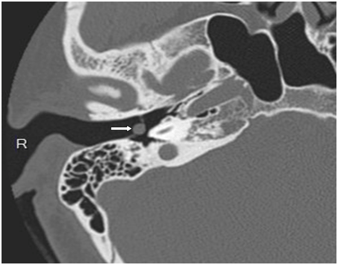
A 45-year-old female complained of right hearing loss with fullness in recent months. She underwent right tympanoplasty type I about 4 years ago. On physical and otoscopic examination, a pearl-like mass about 3 x 4 mm in size over central part of right ear drum was noted (Figure 1A). Pure tone audiometry test showed 30 dB average hearing loss of right ear, and 15 dB of left ear. Tympanogram test showed bilateral type A. Computed tomography with thin cuts of the temporal bone revealed a 3 x 4 mm soft tissue mass over central part of right ear drum (Figure 1B). Excision of the mass under microscope was smoothly done. A cholesteatoma was confirmed by pathology (Figure 2). She was uneventful during a regular follow-up of out-patient department for 12 months.
Figure 1A: A pearl-like mass (arrow) about 3 x 4 mm in size over central part of right ear drum was noted by otoscopic examination.
Figure 1B: Computed tomography (axial view) with thin cuts of the temporal bone revealed a 3 x 4 mm soft tissue mass (arrow) over central part of right ear drum.
Figure 2: Pathology revealed keratin material content; it was compatible with cholesteatoma (Hematoxylin & Eosin staining, 200X).
Discussion
The origin of cholesteatoma or epidermoid cyst is from progressive desquamation of the epithelium. It is a benign collection of keratinized squamous epithelium within the middle ear. Cholesteatomas usually occurred in middle ear cavity or mastoid region, sometimes in external auditory canal. Tympanic membrane cholesteatomas were seldom reported. They could be classified into congenital or acquired according to the pathogenesis [4, 5]. In this case, it was concluded it might be due to previous tympanoplasty and we believed it was acquired. Otalgia, malodorous otorrhea, and hearing loss are the most frequent presenting symptoms regarding cholesteatomas. This patient had mild conductive hearing loss. It is multifactorial regarding to the pathophysiology of the acquired cholesteatoma. Many theories have been recommended and researched on. There are many factors likely to initiate acquired cholesteatoma pathogenesis [6]. Non-iatrogenic or iatrogenic tympanic membrane trauma such as perforation, retraction, displacement, tympanic cavity mucosa disease, ear infection, tympanic membrane disease, and Eustachian tube dysfunction should be taken into account. This case was probably iatrogenic. There are four theories regarding to the pathogenesis of the acquired cholesteatoma. They are (1) retraction pocket cholesteatoma (invagination of the tympanic membrane), (2) basal cell hyperplasia, (3) the migration theory (epithelial in-growth through perforation), and (4) squamous metaplasia of middle ear epithelium [7]. A retraction pocket cholesteatoma is the widely accepted pathogenesis of the acquired type. The theory that negative pressure causes a deepening retraction pocket that, when obstructed, desquamated keratin cannot be cleared from the recess and cholesteatoma happens. The source of the retracted pocket cholesteatomas is believed to be the dysfunction of the Eustachian tube or otitis media with effusion with resultant negative middle ear pressure. In this case, The Eustachian tube function was good. There was no retracted pocket. The migration theory is account for the cause of this case. Normally, the pars flaccida, being less fibrous and less resistant to displacement, is the source of the cholesteatoma. In this case; cholesteatoma was located at the central part of tympanic membrane. The current case concurred with the migration theory. Chronic inflammation appears to play an essential part in multiple etiopathogenic mechanisms of the acquired cholesteatoma. Using of common imaging modalities such as high-resolution computed tomography (CT) scan and magnetic resonance imaging (MRI) are essential to detect and also measure the size of cholesteatomas. It is important for planning a surgical intervention. MRI can differentiate cholesteatomas from other soft tissue masses such as schwannomas, neuromas, or a metastatic mass. CT scans can confirm the existence and offer a suggestion on the size and magnitude of the cholesteatoma. Excision of cholesteatoma is the treatment modality for such disease. Currently, there are no medical treatments that have been demonstrated effective for the treatment of cholesteatomas. Only the treatment of the secondary infectious processes in the cases of acquires cholesteatoma require medical assistance. The microscopic approach enables the surgeon to visualize whole ear drum and hence avoid injuring other part during the operation. Surgical approach to the cholesteatomas is depending on the location, the size and the state of hearing. Most patients operated on had good post-operative recovery as reported in the previous reports. This patient was uneventful during a regular follow-up of out-patient department.
Conclusions
This was a rare occurrence of cholesteatoma. There was no literature had reviewed a case of tympanic membrane cholesteatoma. A congenital or acquired cholesteatoma might grow silently over many years. Excision of tympanic membrane cholesteatoma is the treatment modality for such disease.
Informed Consent
Written informed consent could not be taken from the patient, because of the lack of communication. This case report does not contain any identity information about the patient.
Author Contributions
Concept – H-W Wang; Writing Manuscript – H-W Wang.
Declaration of Interest
The author has no conflict of interest to declare.
Funding
The author declared that this study has received no financial support.
Article Info
Article Type
Case Report & Review of LiteraturePublication history
Received: Mon 21, Oct 2019Accepted: Thu 14, Nov 2019
Published: Fri 29, Nov 2019
Copyright
© 2023 Hsing-Won Wang. This is an open-access article distributed under the terms of the Creative Commons Attribution License, which permits unrestricted use, distribution, and reproduction in any medium, provided the original author and source are credited. Hosting by Science Repository.DOI: 10.31487/j.SCR.2019.05.23
Author Info
Fei-Peng Lee Pin-Zhir Chao Hsing-Won Wang
Corresponding Author
Hsing-Won WangThe Graduate Institute of Clinical Medicine and Department of Otolaryngology, College of Medicine, Taipei Medical University–Shuang Ho Hospital, Taipei, Taiwan, Republic of China
Figures & Tables


References
- Aquino JE, Cruz Filho NA, de Aquino JN (2011) Epidemiology of middle ear and mastoid cholesteatomas: study of 1146 cases. Braz J Otorhinolaryngol 77: 341-347. [Crossref]
- Dähn J, Anschuetz L, Konishi M, Sayles M, Caversaccio M et al. (2017) Endoscopic Ear Surgery for External Auditory Canal Cholesteatoma. Otol Neurotol 38: e34-e40. [Crossref]
- Kong X, Wu H, Ma W, Li Y, Xing B et al. (2016) Cholesteatoma in the sellar region presenting as hypopituitarism and diabetes insipidus. Medicine 95: e2938. [Crossref]
- Richard SA, Qiang L, Lan ZG, Zhang Y, You C (2018) A giant cholesteatoma of the mastoid extending into the foramen magnum: A case report and review of literature. Neurol Int 10: 7625. [Crossref]
- Bennett M, Warren F, Jackson GC, Kaylie D (2006) Congenital cholesteatoma: theories, facts, and 53 patients. Otolaryngol Clin North Am 39: 1081-1094. [Crossref]
- Persaud R, Hajioff D, Trinidade A, Khemani S, Bhattacharyya MN et al. (2007) Evidence-based review of aetiopathogenic theories of congenital and acquired cholesteatoma. J Laryngol Otol 121: 1013-1019. [Crossref]
- Maniu A, Harabagiu O, Perde Schrepler M, Cătană A, Fănuţă B et al. (2014) Molecular biology of cholesteatoma. Rom J Morphol Embryol 55: 7-13. [Crossref]
