Adult Intussusception Secondary to Colorectal Cancer in a Young Woman: A Case Report
A B S T R A C T
Adult intussusception (AI) is uncommon condition that represents 1-5 % of intestinal obstruction and is frequently caused by an underlying disease with 70-90% of cases having a demonstrable cause based on imaging findings and surgical results. The most common causes of colonic AI are neoplasm. We report a case of right colo-colic intussusception sustained by a malignant tumor.
Keywords
Intussusception, bowel obstruction, colon cancer
Introduction
The Colonic intussusceptions is a rare event that deserves a greater degree of interest especially if it occurs in the adult population [1]. Colonic intussusception is caused by a malignancy more frequent respect to small-bowel intussusception, because of the greater prevalence of malignant tumors in the colon than in the small bowel [2, 3]. This condition in adult needs to be thoroughly investigated and certainly deserves surgical treatment [4-6]. We describe below a case of a young woman with no familiarity with colon adenocarcinoma who came to our observation for intestinal obstruction due to intussusception of the large intestine, undergoing laparoscopic right hemicolectomy surgery. Histological examination confirmed the malignant nature of the lesion that caused the obstruction.
Case Report
A 44-year-old woman with no family history of colon adenocarcinoma was brought to our emergency department with a 3-month history of intermittent abdominal pain accompanied by nausea and vomiting. For about 24 hours, symptoms worsened with the presence of fecaloid vomiting. She denied any history of gastrointestinal bleeding, fever, or past abdominal surgery. Her appetite was good but she reported a 6 kg weight loss during the previous 3 months. Clinical examination showed a palpable mass in the right lower quadrant of the abdomen, but digital rectal examination was unremarkable. This mass was firm, non-tender, painful with limited mobility, with signs of peritoneal reaction. Laboratory data showed leukocytosis (17.000/mm3) and PCR was 102 mg/L. An abdominal baseline computed tomography (CT) was carried out. This showed a three-layered structure giving the characteristic target-shaped appearance in the ascending colon, highly suggestive for an ileocolic intussusception (Figures 1-3) sustained by colonic malignant lesion. To avoid the risk of bowel perforation, which is very high in this type of patient, we do not practice the non-operative reduction.

Figure 1: Axial images demonstrate a mass that occupies the epigastrium with the characteristic configuration of “bowel-within-bowel”, in which the layers of the bowel are duplicated forming concentric rings. Mesentery, consisting of fat, vessels and lymph nodes, forms a crescent around the compressed innermost lumen, surrounded by the two layers of the outer enveloping bowel.
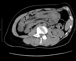
Figure 2: Axial scans acquired caudally demonstrates an extension of the intussusception of the mesogastrium, right flank and right iliac fossa, with involvement of small bowel’s loops, cecum and ascending colon.

Figure 3: On the sagittal plane the intussuscepted bowel presents an irregular wall thickening. This findings is highly suspicious of colonic mass that could act as leading point.
Patient was planned for laparoscopic exploration and eventually definitive surgery. Intra-operatively, we found a colo-colic intussusception with thickening of the colic wall, last intestinal loop attracted and slight proximal intestinal dilation. In addition, multiple lymphadenopathies along the ileocecal artery, but no signs of intestinal ischaemia or peritoneal carcinosis were noted. Consequently, we performed a laparoscopic right hemicolectomy following strict oncologic principles with “en bloc resection” and lymphadenectomy given the risk of an underlying malignancy. Considering this risk, previous reduction of the invaginated segments was not attempted. The specimen was exteriorized through a 5-cm incision in the right upper quadrant, and primary extracorporeal anastomosis was performed using manual sutures. Macroscopic examination revealed a tumor mass of the caecal wall measuring 3×4×3 cm and occupying more than 3-quarters of the circumference with multiple lymphadenopathies along the ileocecal artery were observed (Figure 4). The histological pathological analysis carried out moderately differentiated tubular adenocarcinoma invading the serous (T3) with permeation of the lymphatic and venous capillaries. Of removed lymphatic nodes 6/28 were metastatic. The surgical margins were negative for cancer. Postoperative course was uneventful and patient was discharged 6 days after surgery.

Figure 4: Operative piece: right colon with last ileal loop, a demarcation line can be seen above the cecum attracted by the neoplasm in the ascending colon. The last ileal loop was also attracted.
Discussion
Intussusception is defined as the invagination of one segment of the bowel into an immediately adjacent segment. The intussusceptum refers to the proximal segment that invaginates into the distal segment, or the intussuscipiens (recipient segment). Intussusception, more common in the small bowel and rarely involving only the large bowel. The natural history of intussusception starts with a lead point, usually a neoplasm, which arise as a focal area of traction that draws the proximal bowel within the peristalsing distal bowel. Intussusception is more common in the pediatric population than in the adult population and may be associated with anatomic factors or intestinal infections and management in this population generally starts with non-operative reduction of the intussusceptum using air or contrast enemas [6]. AI is rare, it contributes to less than 5% of all cases of bowel obstructions [2, 3]. Adult intussusception less commonly occurs in the colon than in the small bowel and accounts for only 20 to 25% of all intussusceptions in several reported case series or case report [1, 4, 5, 7-13].
The malignant conditions of the colon associated with intussusception are Adenocarcinoma, Lymphoma and Sarcoma [2, 3]. This justifies the surgical approach in the treatment of this pathology [4-6].
The clinical manifestations of intussusception are typically with pain, nausea, vomiting, constipation, gastrointestinal bleeding, change in bowel habits, constipation, or bloating. More rarely we can find a palpable mass. If the obstruction is not resolved, there may be an evolution in intestinal ischaemia, intestinal perforation and the appearance of signs of shock. Laboratory values typically reveal a high number of white blood cells and altered nonspecific inflammatory markers / acute phase reagents [6]. Preoperative diagnosis remains difficult, while whether the intussusception should be reduced, and the extent of resection, remains controversial. The gold standard is represented by computer tomography CT. It provides information on the presence or absence of intussusception disease, on its location, on intestinal segments involved and on their extent of disease, CT shows also complications such as ischaemia and intestinal wall perforation and helps to plain the most appropriate treatment and to avoid surgery when not indicated.
The optimal surgical approach in adult intussusception is also debatable. In few series, malignancy was the cause in 65% of intussusceptions [14]. Thus, a controversy continues to focus on whether AI should be surgically resected without an attempt at reduction, for fear that undue operative manipulation of a malignant lesion may result in tumor dissemination [15]. We suggest surgically treating adult patients with colonic intussusception due to the high probability of treating a malignant neoplasm and making an oncologically adequate resection. Clinical and instrumental signs of intestinal ischaemia are mandatory for surgery.
Conclusion
When the colon is involved by AI with malignancy is mandatory an immediate surgical treatment to avoid complication such as ischaemia and perforation.
Acknowledgements
Not Applicable.
Author Contributions
D.G., A.D.C., O.T., G.L., V.M., L.B., F.L. and M.T. conceptualized and designed the study, acquired, and analysed data, interpreted the study results, drafted the manuscript, and critically revised the final version of the manuscript.
Funding
None.
Ethics Approval and Consent to Participate
All procedures performed in studies involving human participants were in accordance with the ethical standards of the institutional and/or national research committee and with the 1964 Helsinki Declaration and its later amendments or comparable ethical standards.
Consent
Written informed consent was obtained from the patient for publication of this case report and any accompanying images.
Competing Interests
None.
Article Info
Article Type
Case ReportPublication history
Received: Thu 12, Nov 2020Accepted: Wed 02, Dec 2020
Published: Tue 15, Dec 2020
Copyright
© 2023 Denise Gambardella. This is an open-access article distributed under the terms of the Creative Commons Attribution License, which permits unrestricted use, distribution, and reproduction in any medium, provided the original author and source are credited. Hosting by Science Repository.DOI: 10.31487/j.GSCR.2020.02.10
Figures & Tables
References
- Begos DG, Sandor A, Modlin IM (1997) The diagnosis and management of adult intussusception. Am J Surg 173: 88-94. [Crossref]
- Tamburrini S, Bertucci B, Barresi D (2004) Adult colocolic intussusception: demonstration by conventional MR techniques. Abdom Imaging 29: 42-44. [Crossref]
- Choi SH, Han JK, Kim SH, Lee JM, Lee KH et al. (2004) Intussusception in adults: from stomach to rectum. AJR Am J Roentgenol 183: 691-698. [Crossref]
- Gollub MJ (2011) Colonic Intussusception: Clinical and Radiographic Features. AJR Am J Roentgenol 196: W580-W585. [Crossref]
- Al Radaideh AM, Omari HZ, Bani Hani KE (2018) Adult intussusception : A 14-year retrospective study of clinical assessment and computed tomography diagnosis. Acta Gastroenterol Belg 81: 367-372. [Crossref]
- Marsicovetere P, Ivatury SJ, White B, Holubar SD (2017) Intestinal Intussusception: Etiology, Diagnosis and Treatment. Clin Colon Rectal Surg 30: 30-39. [Crossref]
- Azar T, Berger DL (1997) Adult intussusception. Ann Surg 226: 134-138. [Crossref]
- Ongom PA, Lukande RL (2013) Precipitous intussusception with anal protrusion and complete overt rectal prolapse presenting with intestinal obstruction and an associated rectal adenoma in a young man: a case report. BMC Res Notes 6: 401. [Crossref]
- de Mesquita GHA, Carvalho BJ, de Almeida Medeiros KA, Nii F, Martines DR et al. (2019) Intussusception reveals MUTYH-associated polyposis syndrome and colorectal cancer: a case report. BMC Cancer 19: 324. [Crossref]
- McQuade C, Waters PS, O'Brien C, Crowther S, Torreggiani W et al. (2018) Colorectal intussusception secondary to primary rectal melanoma: A novel case report. Int J Surg Case Rep 44: 78-81. [Crossref]
- Toyoda S, Horii K, Okumura S, Mizumura N, Imagawa A et al. (2018) Sigmoid colon cancer with intussusception prolapsing through the anus treated by elective laparoscopic radical surgery. Nihon Shokakibyo Gakkai Zasshi 115: 87-93. [Crossref]
- Kim YW (2016) A case of postoperative spontaneous intussusception after laparoscopic low anterior resection for rectal cancer. Mil Med Res 3: 19. [Crossref]
- Jennifer M, Agarwal SS, Burke P, Mahoney EJ (2012) Adult intussusception secondary to colorectal cancer in a young man: a case report. J Emerg Med 43: 983-986. [Crossref]
- Nagorney DM, Sarr MG, Mcllrath DC (1981) Surgical management of intussusception in the adult. Ann Surg 193: 230-236. [Crossref]
- Ongom PA, Opio CK, Kijjambu SC (2014) Presentation, aetiology and treatment of adult intussusception in a tertiary Sub-Saharan Hospital: a 10-year retrospective study. BMC Gastroenterol 14: 86. [Crossref]