Apocrine Carcinoma Arising in a Sebaceous Nevus of the Scalp in a 24-Year-Old Female
A B S T R A C T
A case of a 24-year-old female with a two-year history of an intermittently crusting lesion of her scalp is presented. The relatively innocuous-appearing sebaceous nevus, present since birth, made a malignant transformation to an apocrine carcinoma, betrayed by intermittent crusting of a portion of the hamartoma. Current management of sebaceous nevus and apocrine carcinoma is discussed.
Keywords
Nevus sebaceous of Jadassohn, apocrine carcinoma, scalp
Introduction
Nevus sebaceous of Jadassohn (NS) is a hamartoma predominately composed of sebaceous glands and is unbiased towards gender or race. The typical appearance is a yellow-orange, hairless plaque most commonly found on the face or scalp, commonly with a verrucous surface [1, 2]. Prevalence is estimated to be approximately 1.3% [3, 4]. There are three distinct and recognizable stages in the life cycle: in infancy, the lesion remains fairly flat; in adolescence, hormonal influence causes them to become raised and warty; in adulthood, growth ceases, but benign and malignant neoplasms can develop [5]. The most common malignancy is basal cell carcinoma [2, 6-8]. Less commonly appendageal tumors can develop [2, 6-8]. The overall incidence of malignant degeneration is estimated at less than 1% [7].
Cutaneous apocrine carcinoma can arise within NS [9]. The reported occurrences within an NS have been sparse and limited to patients in the 7th decade of life or older [10-12]. There has not yet been a case report published of an apocrine carcinoma arising within an NS in a patient this young of an age.
Case Report
A 24-year-old female presented with a lesion of her right parietal scalp that was present since birth. Two years prior to presentation, an area of ulceration appeared on the anterior portion of the lesion, which subsequently healed and reoccurred several times. Her family physician referred her to a plastic surgeon and subsequently a dermatologist. Reassurances were given that this was a benign lesion, and the occasional appearance of an eschar was due to trauma from routine hair care. The patient was not satisfied and sought out a third opinion.
The presenting lesion was on the right parietal scalp. Except for the lesion, the patient had normal hair development, a normal head and neck examination, and specifically no palpable lymphadenopathy. The lesion was slightly irregular with rounded elevations, was overall well-circumscribed, and alopecic. It was 37 mm by 16 mm in size. It was grossly flesh-coloured, with a few areas that were somewhat yellowish. There was a dry eschar on the anterior portion measuring 10 mm in maximum size (Figure 1). The patient denied traumatizing the area.
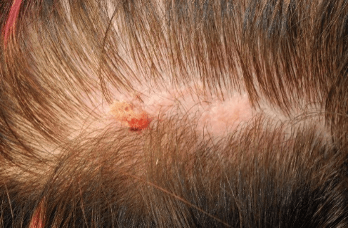
Figure 1: Sebaceous nevus of the scalp with apocrine carcinoma tumor arising in its anterior portion (left of the figure).
An excisional biopsy was carried out with circumferential 3 mm margins. The resultant defect was reconstructed with subgaleal scalp advancement flap closure. The final pathology report indicated that the lesion was an apocrine carcinoma with local infiltration, arising within an NS, with clear resection margins. The metastatic workup was negative. A definitive wide local excision was completed for prophylaxis. The patient remains tumor-free 11 years post-treatment.
Discussion
Cutaneous apocrine carcinoma usually presents as painless, solitary or multiple nodules, with varying colours from red to purple. Ulcerations have been occasionally reported. It is rarely fatal and spreads via lymphatics [9]. Reported occurrences within an NS have been sparse and limited to patients in the 7th decade of life or older [10-12].
Management of NS varies based on the age of the patient, and recommendations are limited to those from expert opinion and case-series studies [1, 2, 13, 14]. From birth to puberty, malignant change is rare, and careful serial observation is appropriate. Any changes in growth patterns or ulceration should prompt a biopsy. After puberty, malignant change becomes more likely. Management should include patient education, and a discussion around close clinical observation versus prophylactic excision, or excision for cosmesis. The rate of malignant transformation in adults is unknown. Estimates from case-series studies are from less than 5% to 30%, with the incidence elevating with age [1, 6].
Conclusion
In summary, the hallmark of malignant change is a change in the characteristics of the NS, as evidenced in the presenting case. Any significant change, including eschar formation and new nodularity, should prompt the clinician to consider incisional or excisional biopsy. This case is unusual as it is the youngest age of presentation reported to date in the literature of degeneration into an apocrine carcinoma and is a reminder to clinicians to investigate changes in an NS regardless of patient age at presentation.
Disclosure
The authors have no financial interest to declare in relation to the content of this article.
Funding
None.
Article Info
Article Type
Case ReportPublication history
Received: Thu 21, May 2020Accepted: Fri 05, Jun 2020
Published: Mon 22, Jun 2020
Copyright
© 2023 Timothy J. Best. This is an open-access article distributed under the terms of the Creative Commons Attribution License, which permits unrestricted use, distribution, and reproduction in any medium, provided the original author and source are credited. Hosting by Science Repository.DOI: 10.31487/j.GSCR.2010.01.01
Figures & Tables
References
- Altaykan A, Ersoy Evans S, Erkin G, Ozkaya O (2008) Basal cell carcinoma arising in nevus sebaceous during childhood. Pediatr Dermatol 25: 616-619. [Crossref]
- Barkham MC, White N, Brundler MA, Richard B, Moss C (2007) Should Naevus Sebaceus Be Excised Prophylactically? A Clinical Audit. J Plas Reconstr Aesthet Surg 60: 1269-1270. [Crossref]
- Edgar N, Schuering RA, Esguerra D, Miller RA, van der Kooi K (2018) Primary Cutaneous Apocrine Carcinoma Arising Within a Nevus Sebaceus. Cutis 102: 291-294. [Crossref]
- Kamyab Hesari K, Balighi K, Afshar N, Aghazadeh N, Rahbar Z et al. (2013) Clinicopathological Study of 1016 Consecutive Adnexal Skin Tumors. Acta Med Iran 51: 879-885. [Crossref]
- Mehregan AH, Pinkus H (1965) Life history of organoid nevi. Special reference to nevus sebaceus of Jadassohn. Arch Dermatol 91: 574-588. [Crossref]
- Domingo J, Helwig EB (1979) Malignant neoplasms associated with nevus sebaceous of Jadassohn. J Am Acad Dermatol 1: 545-556. [Crossref]
- Cribier B, Scrivener Y, Grosshans E (2000) Tumors Arising in Nevus Sebaceus: A Study of 596 Cases. J Am Acad Dermatol 42: 263-268. [Crossref]
- Dalle S, Skowron F, Balme B, Perrot H (2003) Apocrine Carcinoma Developed in Nevus Sebaceus of Jadassohn. Eur J Dermatol 13: 487-489. [Crossref]
- Robson A, Lazar A, Ben Nagi J, Hanby A, Grayson W et al. (2008) Primary Cutaneous Apocrine Carcinoma: A Clinico-Pathologic Analysis of 24 Cases. Am J Surg Pathol 32: 682-690. [Crossref]
- Alessi E, Sala F (1986) Nevus sebaceous. A clinicopathologic study of its evolution. Am J Dermatopathol 8: 27-31. [Crossref]
- Aguayo R, Pallares J, Casanova JM, Baradad M, Sanmartín V et al (2010) Squamous cell carcinoma developing in Jadassohn's sebaceous nevus: case report and review of the literature. Dermatol Surg 36: 1763-1768. [Crossref]
- Paudel U, Jha A, Pokhrel DB, Gurung D, Parajuli S et al (2012) Apocrine Carcinoma Developing in a nevus sebaceous of scalp. Kathmandu Univ Med J 10: 103-105. [Crossref]
- Santibanez Gallerani A, Marshall D, Duarte A, Melnick SJ, Thaller S (2003) Should nevus sebaceous of Jadassohn in children be excised? A study of 757 cases, and literature review. J Craniofacial Surg 14: 658-660. [Crossref]
- Chun K, Vazquez M, Sanchez JL (1995) Nevus sebaceus: clinical outcome and considerations for prophylactic excision. Int J Dermatol 4: 538-541. [Crossref]