Journals
Case Report: An Extreme Case of Alveolar Bone Resorption in an Edentulous Mandible
A B S T R A C T
The alveolar processes of the mandible and maxilla develop in response to tooth eruption and serve as the principal mechanism of tooth support. When teeth are lost or extracted, the result is resorption of the alveolar bone. The rate of resorption is variable between individuals but will progress over time. During dissection, varying examples of alveolar resorption are observed in edentulous donors. A 76-year-old female donor presented an extreme case of alveolar bone resorption during dissection in our Head and Neck Anatomy course. The body of the mandible in this case was extremely short, with a vertical height in the molar region of 5mm and 8mm in the canine area. The dissection revealed the superior surface of the bone to have an open groove containing the inferior alveolar nerve and vessels. The maxilla also demonstrated severe resorption. This loss of bone has significant implications for restoration of function by dental implants or removable prosthesis. It is not unusual to find resorption that has significantly reduced the height of the mandibular body to the extent that the mental foramen opens superiorly on the bone. However, an open mandibular canal with the neurovascular bundle exposed throughout the dental arch is much more unusual. Individuals with this degree of resorption present great challenges in treatment. There is no means to provide retention for a mandibular removable prosthesis. Pressure of prosthesis on the inferior alveolar and mental nerves would potentially produce pain and paresthesia and/or numbness. Placement of dental implants is complicated at best. To provide minimal requirements for implant placement would require vertical augmentation of the mandible along with transposition/reposition of the inferior alveolar nerve
Keywords
Edentulous mandible, residual ridge resorption, dental implants
I N T R O D U C T I O N
The alveolar processes of the mandible and maxilla develop in response to tooth eruption and serve as the principal mechanism of tooth support. When teeth are lost or extracted, the result is resorption of the alveolar bone. The rate of resorption is variable between individuals and is not predictable. However, resorption will certainly progress over time. Studies have shown that age and female gender are risk factors for greater residual mandibular ridge resorption in the posterior mandible [1]. There is no significant association between initial height of the posterior mandibular ridge and the amount of resorption. [1] The number of dentures an individual has worn does not seem to influence the amount of resorption [1]. However, excessive forces in concentrated areas speed resorption in that area and dentures that provide evenly distributed force may result in slower resorption [2]. Osteoporosis does not directly have an impact on the residual ridge height as it effects bone mass and not structure [3]. Because the majority of anatomic donors are of an older age, many encountered in our lab are edentulous. It is common during dissection to encounter varying degrees of alveolar resorption in these donors. This case was the most extreme we have encountered.
Subject
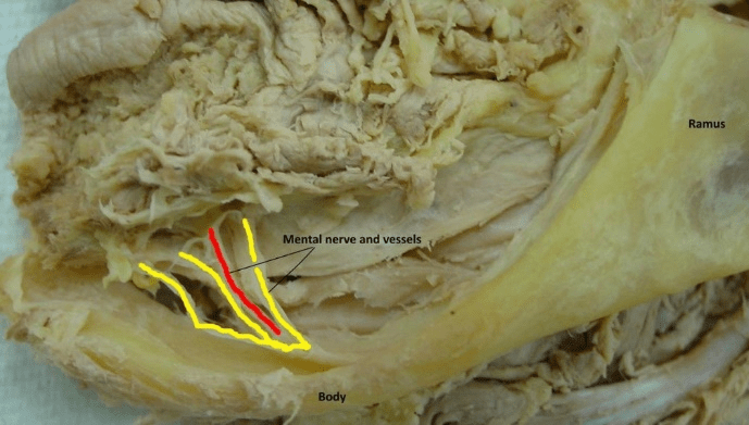
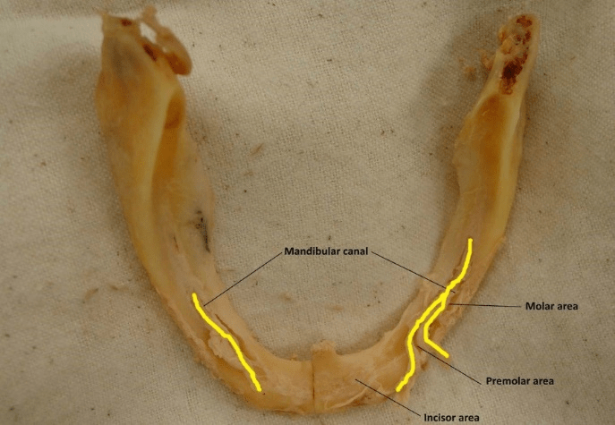
A 76-year-old female donor being studied in the School of Dentistry Head and Neck Anatomy course was found to have an extreme case of alveolar bone resorption (Figure 1).
Table 1: Mandibular measurements of bone thickness
|
|
Right Side |
Left Side |
||
|
Area |
Buccal plate |
Mid bone |
Buccal plate |
Mid bone |
|
Molar |
5.5 |
3.0 |
6.3 |
3.4 |
|
Premolar |
4.0 |
2.3 |
5.0 |
4.0 |
|
Incisor |
6.0 |
7.4 |
6.5 |
6.0 |
Description
During dissection of the oral cavity, the mandible of this donor was found to be extremely delicate. Examination of the mandible revealed that the body of the mandible was extremely narrow, with a vertical height of 5mm in the molar region and 8mm in the canine area along the outer edge of the bone. The depth of the mandible in the central part of the bone, where dental implants would be placed, was less (Table 1). The dissection revealed the superior surface of the bone to have an open groove containing the inferior alveolar nerve and vessels. The neurovascular bundle was exposed throughout the body of the mandible on the left side and had only one area of thin bone covering 6mm in approximately the first molar region on the right. The mental nerves were found branching from the inferior alveolar but there was no mental foramen. The lingual aspect of the bone tapered to a very fine edge that was essentially the attachment of the mylohyoid muscle. The maxilla of this individual also demonstrated severe resorption (Figure 1) and (Figure 2).
Figure 1: Lateral view of the left half of subjects’ mandible prior to removal.
Figure 2: Superior view of subjects’ mandible following removal.
Significance
It is not unusual to find resorption that has significantly reduced the height of the mandibular body to the extent that the mental foramen opens superiorly on the bone. However, an open mandibular canal with the neurovascular bundle exposed throughout the dental arch is more unusual. Pressure of a prosthesis on the inferior alveolar and mental nerves would potentially produce pain and paresthesia and/or numbness. This extreme loss of bone has significant implications for restoration of function by dental implants or removable prosthesis. Individuals with this degree of resorption present great challenges in treatment. [4, 5] With virtually no residual alveolar ridge, there is no means to provide retention for a traditional mandibular removable prosthesis. There is little bone remaining superior to the muscle attachments both on buccal and lingual sides of the bone, which results in very mobile mucosa. Placement of dental implants is complicated at best. [4, 5] The shortest dental implants available in this country are 5-6mm. Typically, in a highly resorbed mandible, two implants would be needed in the canine areas to create a stable base for an overdenture. An overdenture is a removable prosthesis that has tooth or implant support as well as mucosal support. Fracture of the mandible during implant placement is a considerable risk.
In this case, minimal requirements for implant placement are not present. Treatment would require vertical augmentation of the mandible. One treatment method is the grafting of autogenous bone, often from the iliac crest, to the anterior mandible. Anchoring dental implants can be placed through the graft and into the native bone [5]. This may require the transposition/reposition of the inferior alveolar nerve in some cases to provide a field adequate for the procedure. The prosthesis provided must provide broad distribution of force on the bone for the graft to be maintained. Without consistent force, the grafted bone will also resorb. Treatment for a patient such as the one found in this case is complex at best and requires careful diagnostic aids and planning for a successful outcome.
Article Info
Article Type
Case ReportPublication history
Received: Wed 03, Apr 2019Accepted: Wed 17, Apr 2019
Published: Mon 20, May 2019
Copyright
© 2023 Gilbert M. Willett. This is an open-access article distributed under the terms of the Creative Commons Attribution License, which permits unrestricted use, distribution, and reproduction in any medium, provided the original author and source are credited. Hosting by Science Repository.DOI: 10.31487/j.DOBCR.2019.02.03
Author Info
Margaret A. Jergenson Neil S. Norton Barbara J. O’Kane Gilbert M. Willett Laura C. Barritt
Corresponding Author
Gilbert M. WillettDepartment of Oral Biology, Creighton University School of Dentistry, Omaha, NE
Figures & Tables


Table 1: Mandibular measurements of bone thickness
|
|
Right Side |
Left Side |
||
|
Area |
Buccal plate |
Mid bone |
Buccal plate |
Mid bone |
|
Molar |
5.5 |
3.0 |
6.3 |
3.4 |
|
Premolar |
4.0 |
2.3 |
5.0 |
4.0 |
|
Incisor |
6.0 |
7.4 |
6.5 |
6.0 |
References
- Kordatzis K, Wright PS, Meijer HJ (2003) Posterior mandibular residual ridge resorption in patients with conventional dentures and implant overdentures. Int J Oral Maxillofac Implants 18: 447-452. [Crossref]
- Khuder T, Yunus N, Sulaiman E, Ibrahim N, Khalid T et al. (2017) Association between occlusal force distribution in implant overdenture prostheses and residual ridge resorption. J Oral Rehabil 44: 398-404. [Crossref]
- Springe B, Slaidina A, Soboleva U, Lejnieks A (2014) Bone mineral density and mandibular residual ridge resorption. Int J Prosthodont 27: 270-276. [Crossref]
- Sarra Jawad, Craig Barclay, William Whittaker, Martin Tickle, Tanya Walsh (2017) A pilot randomized controlled trial evaluating mini and conventional implant retained dentures on the function and quality of life of patients with an edentulous mandible. BMC Oral Health 17: 53. [Crossref]
- Kuoppala R, Kainulainen VT, Korpi JT, Sandor GK, Oikarinen KS et al. (2013) Outcome of treatment of implant-retained overdenture in patients with extreme mandibular bone resorption treated with bone grafts using a modified tent pole technique. J Oral Maxillofac Surg 71: 1843-1851. [Crossref]
