Journals
Detection of Radioactive Cesium on Granular Particle using Autoradiography, Germanium detector and Energy dispersive X-ray spectrometry
A B S T R A C T
Radioactive Cesium (Cs*) on granular particles were detected using autoradiography, Germanium detector and energy dispersive X-ray spectrometry. The granular particle was included in the suspension of the rainwater tank, which was collected 20 months after the explosion of the Fukushima Dai-ichi Nuclear Power Plant (FDNPP) triggered by the earthquake and following Tsunami on 11 March 2011. The particles are a compound of the silicon oxide and compound of the silicon oxide with Aluminum and Magnesium, respectively. Particle shape was not round but roughly granular with a size of 2μm. The particles are included at the rainwater tank at Ohkuma near FDNPP. The Cs*granular particles are proven to originate to the FDNPP investigating the ratio of Cs137 and Cs134.
K E Y W O R D S
Radioactive cesium, granular particle, autoradiography, germanium detector, energy dispersive X-ray spectrometry, microscopy, FDNPP
I N T R O D U C T I O N
So far, we have succeeded in examining the separation/detection of the radioactive matter that remained in the soil, bamboo and shiitake mushroom as reported in detail in the previous paper [1]. Based on the result, it is concluded that the residual radioactive Cs134 and Cs137 (Cs*), discharged from the FDNPP in the environment and remained on the litter or soils, are mostly water-insoluble. This knowledge agrees with other works: a microscopy based direct observation of Cs* originated to FDNPP was reported, in aerosol particles, called micro-bearing particles with a size of 20μm [2]. Uranium was also reported in the particle as well as some heavy elements later [3].
Regarding the quantification of signal intensity, measurement of the T2 relaxation time (T2 time) in MR imaging can potentially be used for quantitative evaluation of neuropathy associated with change in fluid dynamics and water content as well as providing sensitive detection of localized abnormal regions adjacent to normal nerve regions [9]. Thus, T2 mapping of nerves may be more sensitive to changes in the chemical composition of the nerve tissue associated with CTS than is morphological MR imaging. The clinical usefulness of T2 mapping has been shown in assessments of the articular cartilage, spinal intervertebral disc and Achilles tendon [10-12]. Although T2 mapping of the median nerve in the axial plane has been reported before, the one in the sagittal direction, which is considered more helpful to localize the most severely damaged nerve region, has never been investigated [13].
The amount of the Cs* discharged from the nuclear power station into the environment is estimated to be 1016 Bq. Given that they fell evenly on the ground with a diameter of 50 km, the amount per 1 m2 is 107 Bq [4]. Considering that the number of atoms included in 1 mole Cs*(132g) is 6x10²³, the amount of Cs* that fell on the ground at the explosion of the FDNPP is estimated to be 20x10-7 ng. Cs* flew with air current and dropped on the ground as well as pond and reservoir, too. In the stand-alone pond, which has no outlet for water, Cs* will remain there.
The fact to be concerned is how Cs* has existed in the reservoir, ca, rain water tank, pond and so on, in another word, in the open system without flow path like a river. Because the environment may be remained as it was. Often the water in the pond will be utilized for agriculture. The Cs* granular particle study in the pond or the rain tank was not seen in the literature. We separated and extracted the Cs* that remained in the storage tank of rainwater in the open air. The function of the rainwater tank is similar to the pond but not so large with the size of a several meters square, which is open system without outlet path like a river. The investigation is conducted using autoradiography, Germanium semiconductor radiation detector, and energy dispersive X-ray spectrometry as well as a microscopy work.
Experimental
A 100-cc sample of water from the rainwater tank, collected in Ohkuma Town a several km away from FDNPP in Nov. 2012, were prepared. A pipette of 0.2 mL rainwater including the suspended particles that had submerged on the bottom of the container was sucked. Totally 15 droplets of the above water were dropped on the carbon tapes with both sides adhesive, bonded on Tungsten metal plate in 5 cm square and 0.03 mm thickness. Natural evaporation in laboratory room makes residue on the carbon tape. After it dried, there remained in each sample circular marks with a diameter of several mm; it is called residue in water. Then autoradiography of these samples was processed to investigate whether the radioactive substances are included or not.
Sandwiched structure consisting of two sheets of Imaging Plate (BAS-SR 2040, 200 mm wide x 400 mm long, and the Tungsten metal plate with the residue in water between them is to be exposed to the irradiation by residual Cs*. Narrow space between the imaging plate (IP) sheets and the residue in water was created by a 1 mm-diameter wire on the samples, which could avoid a direct contact IP sheets, and in order to keep a constant distance between the sample and cover plate. After irradiation in special irradiation house made of Lead, IP sheets were read using the special reader system (Fuji Film BAS-2500) with a space resolution of 50μm. Sample preparation flow in the experimental was shown in Fig.2.
For confirming radioactive Cesium, Germanium semiconductor radiation detection was conducted. A rainwater droplet including the suspended particles was dropped on the carbon tape with both sides adhesive, which was bonded on an aluminum desk with a size of 30mm in diameter and 10mm in thickness. Again, a rainwater droplet is dried in air, and then measured using Germanium semiconductor radiation detection. And further, a microscopy work is carried in order to detect X-ray of cesium. Germanium semiconductor radiation detector, CANBERRA GC4020: Energy resolution at 1.33 MeV is smaller than 2.0 keV, was used to identify the radioactive Cesium. A scanning electron microscope, JSM-6010PLUS-LA, was used for energy dispersive X-ray spectrometry.
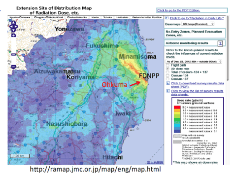
Figure 1: Location of Ohkuma and FDNPP
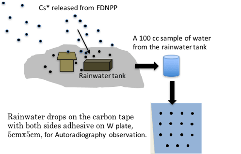
Figure 2: Sample preparation flow in autoradiography experiment. After irradiation in the lead house, IP sheet is read by the special reader system (Fuji Film BAS-2500) with a space resolution of 50μm.
Results and Discussion
Figure 3 shows the result of autoradiography of imaging plate irradiated from droplets sample residue after drying. It was quite obvious that the all droplets contained the radioactive substance. The order of intensity level is shown by red(strong), yellow(medium) and green/blue(weak). Here, the size of the IP image can be considered proportional to the intensity of the radioactive sources contained in the droplets, but it does not equal to the size of substance itself.
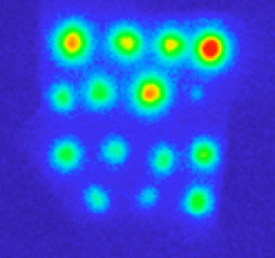
Figure 3: Autoradiography of imaging plate irradiated from droplets sample. The order of intensity level is shown by red(strong), yellow(medium) and green/blue(weak). A size of each image is proportional an intensity of radiation. View size is 5x5cm square.
Figure 4: Shows the results of Germanium semiconductor radiation detection a rainwater droplet including the suspended particles. Measuring sample was located at the center of Al desk with the carbon tape.
Gamma rays were detected for Cs134 at 604.66 keV and 795.76 keV, and for Cs137 at 661.64 keV, respectively. The result verified the existence of radioactive Cesium in the rainwater droplet. Then the sample is investigated to find Cesium in it using a microscopy.
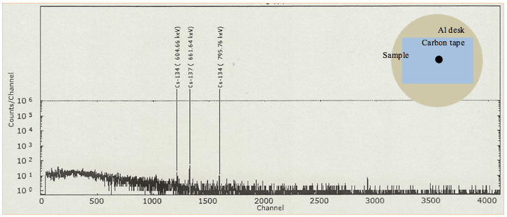
Figure 4: Gamma spectrum from residue particles after natural evaporation of rainwater droplet sample on the carbon tape and aluminum desk. Gamma rays were detected for Cs134 at 604.66 keV and 795.76 keV, and for Cs137 at 661.64 keV, respectively. Germanium semiconductor radiation detector, CANBERRA GC4020 was used.
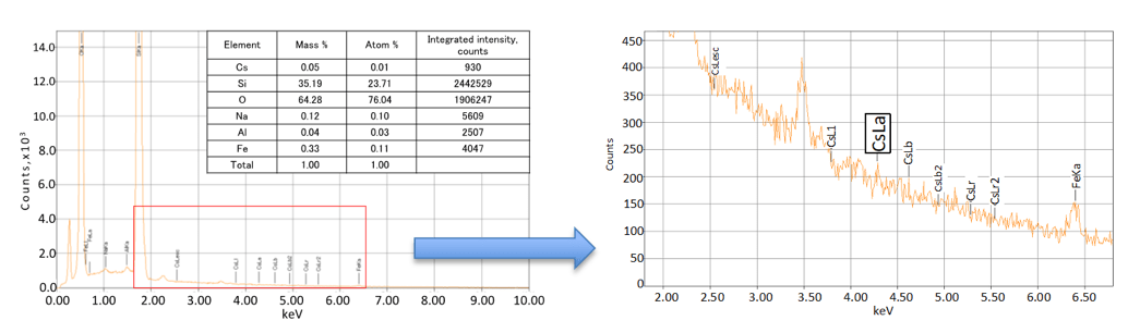
Energy dispersive X-ray spectrometry of the rainwater droplet including the suspended particles detect Cesium on two pieces among the suspended particles. Fig.5 shows a secondary electron-beam image of the particle with a size of 20 micron-meter. The energy spectrum measured at the point 1 is shown. A Cs-La (4.286keV) peak was detected. It includes O, Na, A1, Si, Fe, and Cs with the relative concentration of each being 76.04,0.1,0.03,23.71,0.11, and 0.01(atom%), respectively. The relative density of the number of atoms of Cs was 10-4. This concentration is larger than 1ppm and the value is above the lowest detectable limit, 10-6. Cs peak was also detected in the other four points (2, 3, 4 and 5) mapping measurement, but not in the other particles shown in this secondary electron-beam image. Judging from the information by Germanium semiconductor radiation detector and energy dispersive X-ray spectrometry, radioactive Cs adhered near surface the silicon oxide. The size is 20μm and a shape is not round.
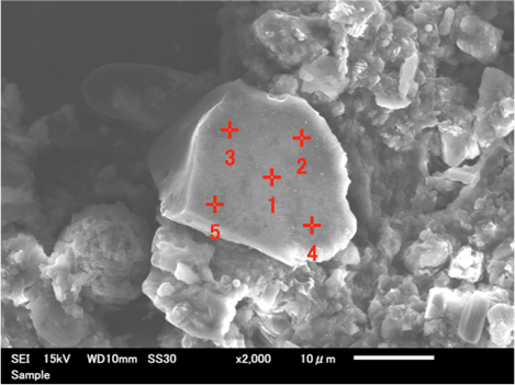
Figure 5: A secondary electron microscopy image of the sample, top, and energy dispersive X-ray spectrometry result of the particle, bottom, for the case of point 1. Plot in the left bottom was partly enlarged for detail, right, bottom. Cs-La peak (4.286keV) was detected in all points from 1 to 5. It includes O, Na, A1, Si, Fe, and Cs and seems to be a silicon oxide.
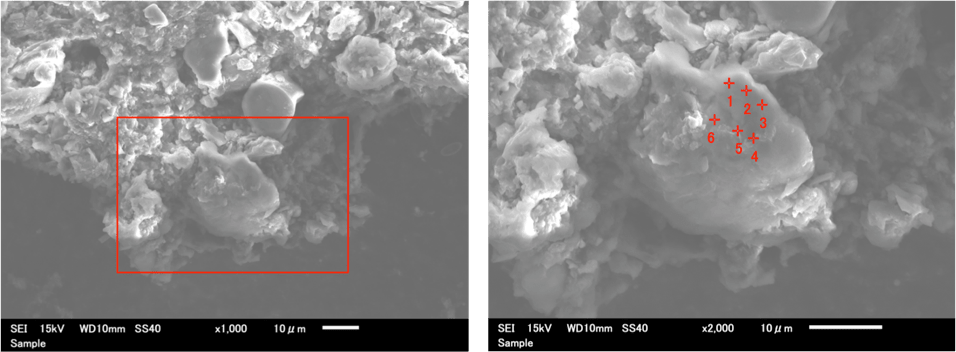
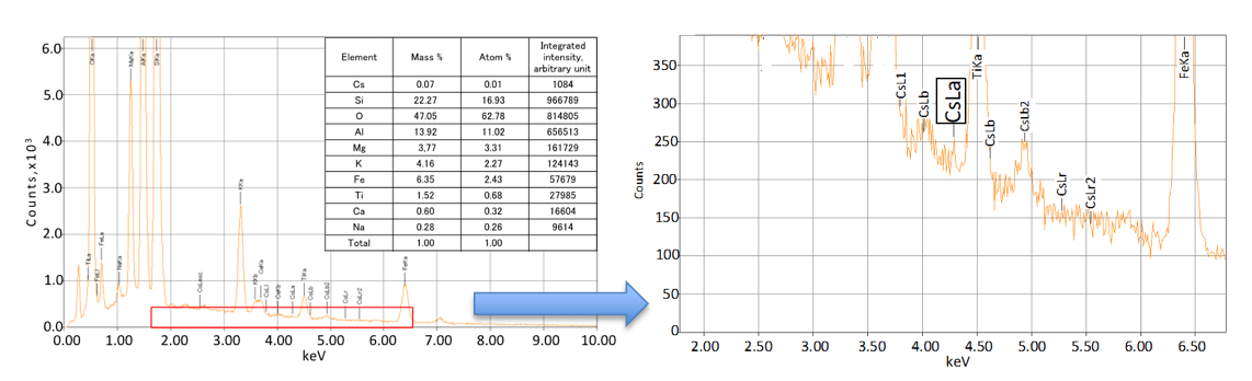
Figure 6: A secondary electron microscopy image of the sample, top, and energy dispersive X-ray spectrometry result of the particle, bottom, for the case of point 1. Image of top in the side is enlarged by square to the right one. Plot in the left bottom was partly enlarged for detail, right, bottom. Cs-La peak(4.286keV) was detected in all points from 1 to 6. It includes Si, O, A1, Mg, K, Fe, Ti, Ca, Na and Cs, and seems to be a compound of silicon oxide with Aluminum, and Magnesium.
Figure 6 shows a secondary electron-beam image of the particle, different from the particle shown in Fig.5, with a size of 20 micron-meter. The energy spectrum measured at the point 1 is shown. A Cs-La (4.286keV) peak was detected. In this case Cs-La was detected near Ti-Ka peak. Often Cs-La peak was hidden in the foot of Ti-Ka peak, but successfully detected due to the reduction of back ground noise.
All of six points analyses shows Cs-La peaks. It includes Si, O, A1, Mg, K, Fe, Ti, Ca, Na and Cs with the relative concentration of each being 16.93,62.78,11.02,3.31,2.27, 2.43, 0.68, 0.32, 0.26 and 0.01(atom%), respectively. Difference from the first particle was that the second particle includes Mg, K and Ti, too. The particle may be a compound of silicon oxide with Aluminum, and Magnesium. Potassium may be originated to plant grown in the rainwater tank.
Hitherto, Ohkura et al. monitored gamma-ray dose rate at monitoring site in Tokai 110km away from FDNPP and quantified radionuclides of Cs-134, 136, 137 and so on, continuously. Most of the ratio of atmospheric concentration of Cs134 to that of Cs137 was found between 0.9 and 1. Ohkura et al., In this work we investigated the water in the rainwater tank near FDNPP and detected radioactive cesium adhered to the particle 20μm in size and shape is not round [5]. In order to check the Cs* is originated to FDNPP or not, the investigation of ratio of Cs134 and Cs137 was done. We stirred contaminated water with suspended particles in the container, collected 33 g out of it and put it in the plastic container, with a nominal volume of 100cm3. Using a TS-100B Becquerel monitor, TECHNO AP, the amount of Cs137 was 1663.1 ± 168.0 Bq/kg, and that of Cs134 was 446.2 ± 93.5 Bq/kg. The ratio of Cs134:Cs137 is 0.27: 1 (measured April 4, 2014). This value can be modified to 0.9: 1 at March 12, 2011. This value validates that Cs* is originated to FDNPP as shown by Ohkura [5].
The information of physical properties, structure and shape was studied.
Water-insoluble Cs*, using autoradiography techniques, in the soil sampled at a primary school near FDNPP, bamboo and soil at Nihonmatsu 30km away from FDNPP, and shiitake mushroom at Yokokawa, 80km away from FDNPP [1].
Air dust samples, captured at Tsukuba 170km away from FDNPP in April - May 2011, were collected, and emitted gamma rays were decided by Ge-detector. A size distribution was determined using a low-pressure cascade impactor, and the results showed the accumulation in the size range between 0.1 and 2μm without particle images taken by microscopy [6].
Micro-bearing particles were reported to include radioactive Cs, using scanning electron microscope with energy dispersion X-ray analyses. Detail analysis was reported then after using multiple synchrotrons radiation-induced X‑ray [2]. The particles were captured in the aerosol in March 2011, at Tsukuba, 174km away from FDNPP [3]. Particles size is between 1-2μm in diameter and concluded to be originated to FDNPP due to co-existence with Uranium and other heavy metals, Ba, Sn, Mo, Zn, and Fe.
Here it is reported that the Cs* granular particle is detected from the rainwater tank, collected in Ohkuma Town a several km away from FDNPP.
Conclusions
- Radioactive cesium on the particle of compound of the silicon oxide and compound of the silicon oxide with Aluminum and Magnesium was detected using autoradiography, Germanium detector and energy dispersive X-ray spectrometry. They existed in the suspended particles of the rainwater storage tank located a several km away from FDNPP with a size of 20 μm. The particle shape is not round but granular.
- The ratio of Cs137 and Cs134 is 0.9: 1 at March 12, 2011. The value validates that radioactive cesium is originated to FDNPP.
Article Info
Article Type
Research ArticlePublication history
Received: Wed 02, May 2018Accepted: Mon 14, May 2018
Published: Fri 25, May 2018
Copyright
© 2023 Kenji Kikuchi. This is an open-access article distributed under the terms of the Creative Commons Attribution License, which permits unrestricted use, distribution, and reproduction in any medium, provided the original author and source are credited. Hosting by Science Repository.DOI: 10.31487/j.RDI.2018.10.004
Author Info
Corresponding Author
Kenji KikuchiFrontier Research Center for Applied Atomic Sciences, Ibaraki University, 162-1 Shirakata, Tokai, Naka, Ibaraki 316-1106, Japan
Figures & Tables
References
1. Nobuo Niimura, Kenji Kikuchi, Ninh Duc Tuyen, Masakazu Komatsuzaki, Yoshinobu Motohashi (2015) Physical properties, structure, and shape of radioactive Cs from the Fukushima Daiichi Nuclear Power Plant accident derived from soil, bamboo and shiitake mushroom measurements. J EnvironRadioact 139: 234-239. DOI.org/10.1016/j.jenvrad.2013.12.020. [Crossref]
2. Kouji Adachi, Mizuo Kajino, Yuji Zaizen, Yasuhito Igarashi, (2013) Emission of spherical cesium-bearing particles from an early stage of the Fukushima nuclear accident. Sci Rep 3:2554. [Crossref]
3. Yoshinari Abe, Yushin Iizawa, Yasuko Terada, Kouji Adachi, Yasuhito Igarashi, et al. (2014) analytical chemistry 86: 8521-8525.
4. Masamichi CHINO, Hiromasa NAKAYAMA, Haruyasu NAGAI, Hiroaki TERADA, Genki KATATA, et al. (2011) Preliminary Estimation of Release Amounts of 131I and 137Cs Accidentally Discharged from the Fukushima Daiichi Nuclear Power Plant into the Atmosphere. J nuclear sci tech 48: 1129-1134 DOI.org/10.1080/18811248.2011.9711799.
5. Takehisa OHKURA, Tetsuya OISHI, Mitsumasa TAKI, Yukio SHIBANUMA Masamitsu KIKUCHI, Hitoshi AKINO, et al. (2012) Emergency Monitoring of Environmental Radiation and Atmospheric Radionuclides at Nuclear Science Research Institute, JAEA Following the Accident of Fukushima Daiichi Nuclear Power Plant, JAEA-Data / Code.
6. Naoki Kaneyasu, Hideo Ohashi, Fumie Suzuki, Tomoaki Okuda, Fumikazu Ikemori (2012) Sulfate Aerosol as a Potential Transport Medium of Radiocesium from the Fukushima Nuclear Accident. Environmental Sci Tech 46: 5720-5726.