Journals
Flow, Filling Ability and Apical Extrusion of New Calcium Silicate-Based Sealers: A Micro-Computed Tomographic Study
A B S T R A C T
Aim: To evaluate flow, filling ability, and apical extrusion of three calcium silicate-based sealers, Neo MTA Plus (Avalon, USA), Bio-C Sealer (Angelus, Brazil) and Sealer Plus BC (MK Life, Brazil), in comparation with the gold standard based on epoxy resin, AH Plus (Dentsply, Germany).
Methodology: The flow was evaluated based on ISO 6876/2012 standard (mm) and in area (mm2). For assessment of filling ability and apical extrusion of the sealers, curved artificial canals were prepared up to size 25.06, and filled with the sealers by the single cone technique. The samples were scanned in micro-computed tomography (SkyScan 1272. Bruker, Belgium) after preparation and after filling of the root canals. The percentage of voids throughout the entire extension of the root canals and in the apical third, besides the volume of apical extrusion of each sealer were calculated. Data were statistically analyzed using ANOVA/Tukey tests (α = 0.05).
Results: Bio-C Sealer showed the highest flow and NeoMTA Plus had the lowest value (p < 0.05). Bio-C Sealer had lower percentage of total voids than NeoMTA Plus (p < 0.05). AH Plus had greater percentage of voids in the apical third than Bio-C Sealer and Sealer Plus BC (p < 0.05). Bio-C Sealer showed higher volume of extrusion (p < 0.05) than AH Plus and NeoMTA Plus.
Conclusion: Bio-C Sealer and Sealer Plus BC had greater flow and proper filling ability in the apical third. However, these sealers presented high volume of apical extrusion. NeoMTA Plus provided less sealer extrusion, low flow, and more presence of voids.
Keywords
Dental materials, endodontics, physical properties, root canal sealer, x-ray microtomography
Introduction
Root canal treatment is performed aiming to save teeth [1]. Since a three-dimensional root canal sealing is essential for the success of endodontic therapy, root canal sealers should present flow, aiming to fill the anatomical complexities of the root canal system [2, 3]. However, excessive flow can lead to material extrusion, which may have an impact on the periapical repair process [3]. Therefore, flow and filling of endodontic materials should be evaluated. Micro-computed tomography (micro-CT) is performed to evaluate the filling in obturated root canals [4]. New methodologies using micro-CT can also be applied to complement the conventional flow test stablished by ISO 6876 standard [5].
AH Plus (Dentsply DeTrey GmbH, Konstanz, Germany) is an endodontic sealer mainly composed of epoxy resin, which is considered as gold standard regarding physicochemical properties [6]. On the other hand, calcium silicate-based endodontic sealers has been highlighted, due to their biocompatibility and bioactivity, stimulating mineralization [7-9]. Neo MTA Plus (Avalon Biomedic Inc. Bradenton, FL, USA) is a tricalcium silicate-based material, which can be used as reparative material or root canal sealer, according to its powder-to-gel ratio. NeoMTA Plus presents biocompatibility and promotes induction of mineralized tissue [9-12]. Although this sealer shows penetration into the dentinal tubules, there is no study evaluating its physicochemical properties when used as a root canal sealer [13].
Premixed, ready-to-use calcium silicate-based endodontic sealers have been developed for root canal obturation [14]. Recently, the new calcium silicate-based Sealer Plus BC (MK Life, Porto Alegre, RS, Brazil) and Bio-C Sealer (Angelus, Londrina, PR, Brazil) were introduced into the market. Sealer Plus BC and Bio-C Sealer are biocompatible and have appropriate setting time, flow, and radiopacity, besides alkalinization capacity [15-18]. Although the solubility of these sealers is higher than the rates required by ISO 6876 standard, both materials showed a decrease in solubility when immersed in phosphate-buffered saline (PBS), in addition to a low volumetric change after immersion in distilled water or PBS [17-19].
Since assessments of physicochemical properties are still necessary to evaluate the impact of calcium silicate-based sealers on the outcome of root canal treatment [7], and considering that up to now, no study was performed using micro-CT to investigate the filling ability and apical volume extrusion of NeoMTA Plus, Bio-C Sealer or Sealer Plus BC, the current research was performed. The null hypothesis was that would be no difference among the different sealers regarding their flow, filling ability and apical extrusion.
Materials and Methods
I Flow Evaluation
The flow test was conducted in accordance with ISO Standard 6876/2012 [20]. After manipulation of the sealers, 0.05 mL of the material was placed in the center of a glass plate by using a syringe (n = 10). At 180 ± 5 seconds after manipulation, another glass plate (20 g) was placed on the plate with the sealer, and a 100-gram weight was put on the top plate and kept for 10 minutes. After this period, the maximum and minimum diameters of the material on the glass plate were measured. When a difference of less than 1 mm between the diameters was observed, the mean value was recorded. A second evaluation was made by photographing the sealer on the plate alongside a millimeter ruler. The images obtained were evaluated using the Image Tool 3.0 software (UTHSCSA, San Antonio, TX) to obtain the area of flow of the sealer expressed in mm², according to Tanomaru-Filho et al. [21].
II Evaluation of Filling Ability and Apical Extrusion
Acrylic resin models with artificial canals (IM Brazil Ltda., São Paulo, SP, Brazil) were used (n = 32). The artificial canals had standard size (18 mm), 60° angle of curvature and 5 mm radius and the center of the curvature were 5 mm from the end of the canal.
The apical patency was determined by inserting a size 10 K-file (Dentsply, Maillefer, Ballaigues, Switzerland) until the apical foramen. Working length (WL) was established at 1 mm short of the end of the canal (WL = 17 mm). A single operator prepared all the specimens using the ProDesign Logic system (Easy Equipamentos Odontológicos, Belo Horizonte, MG, Brazil). The 25/.01 file was used in continuous rotation at 350 rpm speed and 1 Ncm torque, using in-and-out movements up to the working length. Then, the 25/.06 file was used at 600 rpm speed and 4 Ncm torque, using an endodontic motor (VDW Silver, VDW GmbH, Munich, Germany). The canals were irrigated with 2.5 mL of distilled water after each instrument by using a disposable syringe and a 27-G NaviTip needle (Ultradent, South Jordan, UT). The prepared canals were divided in four groups (n = 8). The canals were filled by single cone technique with each sealer. The endodontic sealers and their respective manufacturers, compositions and proportions are described in (Table 1).
Table 1: Root canal sealers, their manufacturers, composition, and proportions used.
|
Sealer |
Manufacturer |
Composition |
Proportion |
|
AH Plus |
Dentsply De Trey GmbH, Konstanz, Germany |
Epoxy resin bisphenol-A and bisphenol-F, calcium tungstate, zirconium oxide, silica, iron pigments. Dibenzyl diamide, Aminoadamantane, silicone oil. |
Equal portions (by length) of the base and catalyst pastes |
|
Neo MTA Plus |
Avalon Biomed Inc, Bradenton, FL, USA |
Powder: tricalcium silicate, dicalcium silicate, tantalum oxide, tricalcium aluminate, and calcium sulphate. Liquid: water-based gel with thickening agent and water-soluble polymers. |
1 scoop of powder to 1 drop of gel (0,33 g : 150 μL) |
|
Sealer Plus BC |
MK Life, Porto Alegre, RS, Brazil |
Zirconium oxide, tricalcium silicate, dicalcium silicate, calcium hydroxide, and propylene glycol. |
Premixed, ready-to-use |
|
Bio-C Sealer |
Angelus, Londrina, PR, Brazil |
Calcium silicates, calcium aluminate, calcium oxide, zirconium oxide, iron oxide, silicon dioxide, dispersing agent. |
Premixed, ready-to-use |
AH Plus and NeoMTA Plus were manipulated in accordance with the manufacturer’s specifications and the sealers were inserted into the canals with a Lentulo spiral #35 (Dentsply Maillefer, Ballaigues, Switzerland). Bio-C Sealer and Sealer Plus BC were injected into the root canal 4 mm short of the WL, using the syringe and plastic needle provided by their manufacturers. Subsequently, 25/.06 gutta-percha cones (Tanari industrial Ltda., São Paulo, Brazil), which were previously selected according to the tip size and taper measured in the profilometer (Profile Projector Nikon Model 6C-2), were inserted into each canal up to the WL. Then, the gutta-percha cones were cut at the cervical level and the remaining material was compacted with a heated plugger. A radiograph was taken to verify the obturation quality. The coronal portion was sealed with a provisional restorative material (Coltosol. Vigodent, Rio de Janeiro, Brazil). All specimens were stored in an oven at 37°C in relative humidity for 7 days to allow the sealers to set completely. For NeoMTA Plus, Bio-C Sealer and Sealer Plus BC, which require moisture for setting, two pieces of wet cloth were placed over the samples, as described by Tanomaru-Filho et al. [22].
III Micro-CT Imaging Analysis
The artificial roots were scanned using micro-CT (SkyScan 1272. Bruker, Kontich, Belgium) after preparation, and after the obturation of the root canals. The roots were positioned in a standardized device, allowing the specimens to remain in the same position at all times during scanning procedures. The scanning parameters were defined by pilot study. After preparation the samples were scanned using the following parameters: 60 kV of power, energy of 166 mA, evolution cycle of 180°, rotation of 0.5, aluminum filter (0,25 mm) and voxel size of 15 µm. After canals filling, the scanning parameter were: 80 kV of power, energy of 125 mA, evolution cycle of 180°, rotation of 0.5, aluminum filter (1,00 mm) and voxel size of 15 µm. The post-preparation and post-obturation images obtained were reconstructed using the NRecon software (v.1.6.10.4, Bruker).
The reconstructed images, obtained before and after obturation, were superimposed by means of geometric alignment in the Data Viewer software (v.1.5.1, Bruker). Quantitative analyses were then performed using the CTAn software (v1.15.4.0, Bruker) by applying task lists, and arithmetic and logic operations between the superimposed sections. Root canal volume, filling material volume (gutta-percha and sealer), and voids volume were quantified. The gray scale range required to recognize each object under study was determined in a density histogram by using a global threshold method. The volume of voids was calculated by subtracting the filling material volume from the post-obturation root canal volume: [volume of voids = volume of canal – volume of filling]. The percentage volume of voids was calculated by using the following formula: [Percentage of voids = (volume of voids X 100)/volume of canal]. Qualitative analyses were performed by means of models obtained by using CTVox software (CTVox v. 3.2, Bruker). Evaluation was performed at all root canal extension and in the apical third. The value of approximately 9 mm was determined for the total length analysis and approximately 3 mm for apical third. A schematic figure showing all the process to micro-CT assessment is presented in (Figure 1).
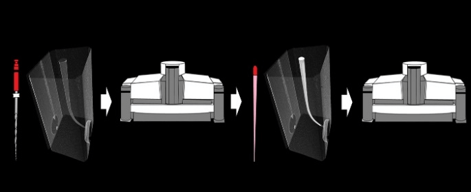
Figure 1: Schematic figure representing the filling ability and apical extrusion assessments in micro-CT. Acrylic resin models with artificial canals were prepared using the ProDesign Logic system and the first scanning was performed in micro-CT SkyScan 1272. The specimens were filled with the different sealers and scanned again.
IV Statistical Analysis
All data were analyzed with statistical software package GraphPad Prism 7.00 (GraphPad Software, La Jolla California USA). Data were submitted to normality test, and then to the parametric one-way analysis of variance (ANOVA) statistical test and to the Tukey multiple comparison test, with significance level at 5%.
Results
Bio-C Sealer had the highest flow (mm and mm²), followed by Sealer Plus BC and AH Plus (p < 0.05). NeoMTA Plus had the lowest value, below 17 mm recommended by ISO 6876:2012 (p < 0.05) [20]. The flow values are represented in (Table 2).
Table 2: Mean and standard deviation of the results of flow (mm and mm²).
|
Sealers/Tests |
AH Plus |
NeoMTA Plus |
Sealer Plus BC |
Bio-C Sealer |
|
Flow (mm) |
21.65 (±0.96)c |
14,38 (±0,72)d |
25.89 (±0.95)b |
31.55 (±1.22)a |
|
Flow (mm²) |
430.2 (±98.37)c |
190,4 (±31,86)d |
718.1 (±53.37)b |
877.2 (±43.92)a |
Different letters indicate statistically significant difference among experimental groups (p < 0.05).
Table 3: Mean and standard deviation of the results of voids (%) and extrusion (mm³) in artificial canals filled by single cone with different endodontic sealers.
|
Sealers/Tests |
AH Plus |
NeoMTA Plus |
SealerPlus BC |
Bio-C Sealer |
|
Total Voids (%) |
0.393 (±0.13)ab |
0.867 (±0.45)a |
0.533 (±0.26)ab |
0.086 (±0.04)b |
|
Apical Voids (%) |
1.636 (±0.48)a |
1.045 (±0.44)ab |
0.885 (±0.23)bc |
0.379 (±0.15)c |
|
Apical Extrusion (mm³) |
0.202 (±0.11)b |
0.518 (±0.21)b |
0.750 (±0.25)ab |
1.165 (±0.45)a |
Different letters indicate statistically significant difference among experimental groups (p < 0.05).
Regarding the micro-CT assessment of filling ability and extrusion volume of the sealers (Table 3 & Figure 2), Bio-C Sealer presented lower percentage of total voids than NeoMTA Plus (p < 0.05). Sealer Plus BC and AH Plus were similar to Bio-C Sealer and NeoMTA Plus (p > 0.05).
When evaluating the apical third, AH Plus had higher percentage of voids than Bio-C Sealer and Sealer Plus BC (p < 0.05). NeoMTA Plus was similar to AH Plus and Sealer Plus BC (p > 0.05). Bio-C Sealer presented lower percentage of voids in apical third than NeoMTA Plus (p < 0.05). Bio-C Sealer had higher extrusion volume than AH Plus and NeoMTA Plus (p < 0.05). Sealer Plus BC was similar to the other sealers (p > 0.05).
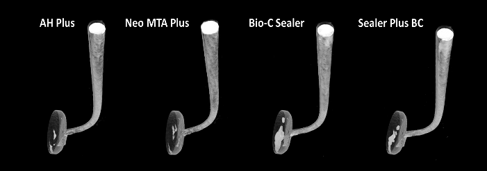
Figure 2 : 3D Models using CTVox software showing the canals filled with AH Plus, Neo MTA Plus, Bio-C Sealer and Sealer Plus BC.
Discussion
The aim of the current study was to evaluate flow, filling capacity and volume of apical extrusion of new premixed ready-to-use bioceramic sealers (Sealer Plus BC and Bio-C Sealer), in comparison with a calcium silicate-based sealer composed of powder and gel (NeoMTA Plus) and an epoxy resin-based material, considered the gold standard regarding physicochemical properties (AH Plus). Differences are detected among the sealers evaluated, rejecting our null hypothesis.
Flow rate of the root canal sealers was evaluated since this property should allow the filling of irregularities, leading to greater penetration into accessory canals, isthmus, and dentinal tubules [6, 23]. In the current study, Bio-C Sealer had the highest flow, followed by Sealer Plus BC, AH Plus and the lowest value was obtained by NeoMTA Plus. There is no study evaluating this characteristic for NeoMTA Plus and no parameters for comparison. However, even did not complying with the ISO standard, a previous study showed a proper tubule penetration for this sealer [13].
The flow may be influenced by the composition and particle size of the materials [24]. Nanoparticles are widely used in dentistry aiming to improve the properties of materials [25]. Despite the presence of resin in AH Plus, which allows a higher flow capacity, the premixed ready-to-use calcium silicate-based sealers showed greater flow than AH Plus, which can be explained by the small size and degree of viscosity of calcium silicate particles [6, 26, 27]. This high flow may justify the three-dimensional filling promoted by Bio-C Sealer and Sealer Plus BC, with low percentage of voids for both sealers.
AH Plus and NeoMTA Plus had similar voids percentage. To date, there are no studies comparing the filling capacity of root canals with NeoMTA Plus, making difficult direct comparisons. However, several studies have shown satisfactory filling capacity for bioceramic sealers, having similar filling ability to AH Plus [28-30]. Additionally, the apical third is considered critical for endodontic treatment success and control of re-infection [31, 32]. Bio-C Sealer and Sealer Plus BC had lower percentage of voids in the apical third than AH Plus.
Although the high flow rate can lead to a lower total voids percentage for Bio-C Sealer in comparison with NeoMTA Plus and also a better filling in the apical third in comparison with NeoMTA Plus and AH Plus, an excessive flow may increase the risk of apical extrusion, which may impair the apical repair process [3, 23]. Moreover, even that the insertion of Bio-C Sealer and Sealer Plus BC in the canals by means of an applicator syringe may have contributed to the better filling of the canals, a greater apical extrusion of these sealers can be a consequence of this technique. Overextension of endodontic materials beyond the apical foramen could damage periodontal tissues, and in posterior lower teeth could damage the inferior alveolar nerve [33, 34]. Therefore, there is a greater chance of a consequent postoperative pain [23]. Nevertheless, a previous study evaluated three hundred seven teeth with an average follow-up time of 30 months after endodontic treatment using a ready-to-use calcium silicate-based sealer (EndoSequence BC Sealer, Brasseler, USA, Savannah, GA) with a single-cone technique [35]. The authors observed sealer extrusion in 47.4% of the cases and concluded that this factor did not have any significant effect on the treatment outcome, which showed an overall success rate of 90.9%.
Furthermore, an important limitation of the current study was the absence of tissues surrounding the resin block, differentiating this in vitro study from a real clinical condition. In addition, most of the studies that evaluated root canal filling used root canals of extracted human teeth, whereas in the present study it was used simulated canals in blocks of acrylic resin [28-30]. Although the artificial canals do not reproduce the characteristics of root dentin, there is a great difficulty in obtaining human teeth with similar conditions for comparison [36]. Therefore, laboratory studies with acrylic blocks and simulated canals are commonly used, which ensure a control of experimental conditions, allowing only the variables of interest to be analyzed [37, 38].
Conclusion
Within the limitations of this in vitro study, we can conclude that the premixed ready-to-use bioceramic sealers, Bio-C Sealer and Sealer Plus BC had a high flow and filling ability in the apical third. However, these sealers presented greater apical extrusion. NeoMTA Plus provided less extrusion, but low flow and more voids.
Conflicts of Interest
None.
Funding
This study was financed in part by the Coordenação de Aperfeiçoamento de Pessoal de Nível Superior - Brasil (CAPES) - Finance Code 001 and was supported by São Paulo Research Foundation – FAPESP (2016/00321-0, 2017/14305-9, 2017/19049-0, and 2018/19665-6).
Author Contributions
Designed the study: Mário Tanomaru-Filho, Juliane Maria Guerreiro-Tanomaru; Executed the study or conducted the experiments: Jáder Camilo Pinto, Fernanda Ferrari Esteves Torres, Pedro Henrique Fiorin de Souza, Mariana Carvalho Pereira; Have taken part in analysing the data: Jáder Camilo Pinto, Fernanda Ferrari Esteves Torres, Pedro Henrique Fiorin de Souza, Mariana Carvalho Pereira; Supported in documenting the article and drawing the conclusion: Jáder Camilo Pinto, Fernanda Ferrari Esteves Torres, Mário Tanomaru-Filho, Juliane Maria Guerreiro-Tanomaru; Spearheaded the project as principal investigators: Mário Tanomaru-Filho, Juliane Maria Guerreiro-Tanomaru.
Article Info
Article Type
Research ArticlePublication history
Received: Thu 21, May 2020Accepted: Sat 06, Jun 2020
Published: Mon 15, Jun 2020
Copyright
© 2023 Mario Tanomaru Filho. This is an open-access article distributed under the terms of the Creative Commons Attribution License, which permits unrestricted use, distribution, and reproduction in any medium, provided the original author and source are credited. Hosting by Science Repository.DOI: 10.31487/j.DOBCR.2020.03.04
Author Info
Corresponding Author
Mario Tanomaru FilhoDepartment of Restorative Dentistry, São Paulo State University (UNESP), School of Dentistry, Araraquara, São Paulo, Brazil
Figures & Tables
Table 1: Root canal sealers, their manufacturers, composition, and proportions used.
|
Sealer |
Manufacturer |
Composition |
Proportion |
|
AH Plus |
Dentsply De Trey GmbH, Konstanz, Germany |
Epoxy resin bisphenol-A and bisphenol-F, calcium tungstate, zirconium oxide, silica, iron pigments. Dibenzyl diamide, Aminoadamantane, silicone oil. |
Equal portions (by length) of the base and catalyst pastes |
|
Neo MTA Plus |
Avalon Biomed Inc, Bradenton, FL, USA |
Powder: tricalcium silicate, dicalcium silicate, tantalum oxide, tricalcium aluminate, and calcium sulphate. Liquid: water-based gel with thickening agent and water-soluble polymers. |
1 scoop of powder to 1 drop of gel (0,33 g : 150 μL) |
|
Sealer Plus BC |
MK Life, Porto Alegre, RS, Brazil |
Zirconium oxide, tricalcium silicate, dicalcium silicate, calcium hydroxide, and propylene glycol. |
Premixed, ready-to-use |
|
Bio-C Sealer |
Angelus, Londrina, PR, Brazil |
Calcium silicates, calcium aluminate, calcium oxide, zirconium oxide, iron oxide, silicon dioxide, dispersing agent. |
Premixed, ready-to-use |
Table 2: Mean and standard deviation of the results of flow (mm and mm²).
|
Sealers/Tests |
AH Plus |
NeoMTA Plus |
Sealer Plus BC |
Bio-C Sealer |
|
Flow (mm) |
21.65 (±0.96)c |
14,38 (±0,72)d |
25.89 (±0.95)b |
31.55 (±1.22)a |
|
Flow (mm²) |
430.2 (±98.37)c |
190,4 (±31,86)d |
718.1 (±53.37)b |
877.2 (±43.92)a |
Different letters indicate statistically significant difference among experimental groups (p < 0.05).
Table 3: Mean and standard deviation of the results of voids (%) and extrusion (mm³) in artificial canals filled by single cone with different endodontic sealers.
|
Sealers/Tests |
AH Plus |
NeoMTA Plus |
SealerPlus BC |
Bio-C Sealer |
|
Total Voids (%) |
0.393 (±0.13)ab |
0.867 (±0.45)a |
0.533 (±0.26)ab |
0.086 (±0.04)b |
|
Apical Voids (%) |
1.636 (±0.48)a |
1.045 (±0.44)ab |
0.885 (±0.23)bc |
0.379 (±0.15)c |
|
Apical Extrusion (mm³) |
0.202 (±0.11)b |
0.518 (±0.21)b |
0.750 (±0.25)ab |
1.165 (±0.45)a |
Different letters indicate statistically significant difference among experimental groups (p < 0.05).
References
- Lokhande PR, Balaguru S (2020) A mathematical model for root canal preparation using Endodontic file. J Oral Biol Craniofac Res 10: 396-400. [Crossref]
- Bhatt A, Rajkumar B (2019) A comparative evaluation of cyclic fatigue resistance for different endodontic NiTi rotary files: An in-vitro study. J Oral Biol Craniofac Res 9: 119-121. [Crossref]
- Duarte MA, Ordinola-Zapata R, Bernardes RA, Bramante CM, Bernardineli N et al. (2010) Influence of calcium hydroxide association on the physical properties of AH Plus. J Endod 36: 1048-1051. [Crossref]
- Lokhande PR, Balaguru S (2020) The quantification of percentage filling of gutta-percha in obturated root canal using image processing and analysis. J Oral Biol Craniofac Res 10: 28-32. [Crossref]
- Tanomaru Filho M, Torres FFE, Bosso Martelo R, Chávez Andrade GM, Bonetti Filho I et al. (2017) A Novel Model for Evaluating the Flow of Endodontic Materials Using Micro-computed Tomography. J Endod 43: 796-800. [Crossref]
- Silva Almeida LH, Moraes RR, Morgental RD, Pappen FG (2017) Are Premixed Calcium Silicate-based Endodontic Sealers Comparable to Conventional Materials? A Systematic Review of In Vitro Studies. J Endod 43: 527-535. [Crossref]
- Donnermeyer D, Burklein S, Dammaschke T, Schafer E (2019) Endodontic sealers based on calcium silicates: a systematic review. Odontology 107: 421-436. [Crossref]
- Gandolfi MG, Siboni F, Botero T, Bossù M, Riccitiello F et al. (2015) Calcium silicate and calcium hydroxide materials for pulp capping: biointeractivity, porosity, solubility and bioactivity of current formulations. J Appl Biomater Funct Mater 13: 43-60. [Crossref]
- Tanomaru Filho M, Andrade AS, Rodrigues EM, Viola KS, Faria G et al. (2017) Biocompatibility and mineralized nodule formation of Neo MTA Plus and an experimental tricalcium silicate cement containing tantalum oxide. Int Endod J 50: e31-e39. [Crossref]
- Pinheiro LS, Iglesias JE, Boijink D, Mestieri LB, Kopper PMP et al. (2018) Cell Viability and Tissue Reaction of NeoMTA Plus: An In Vitro and In Vivo Study. J Endod 44: 1140-1145. [Crossref]
- Walsh RM, Woodmansey KF, He J, Svoboda KK, Primus CM et al. (2018) Histology of NeoMTA Plus and Quick-Set2 in Contact with Pulp and Periradicular Tissues in a Canine Model. J Endod 44: 1389-1395. [Crossref]
- Rodriguez Lozano FJ, Collado Gonzalez M, Lopez Garcia S, García Bernal D, Moraleda J et al. (2019) Evaluation of changes in ion release and biological properties of NeoMTA-Plus and Endocem-MTA exposed to an acidic environment. Int Endod J 52: 1196-1209. [Crossref]
- McMichael GE, Primus CM, Opperman LA (2016) Dentinal Tubule Penetration of Tricalcium Silicate Sealers. J Endod 42: 632-636. [Crossref]
- Bidar M, Sadeghalhoseini N, Forghani M, Attaran N (2014) Effect of the smear layer on apical seals produced by two calcium silicate-based endodontic sealers. J Oral Sci 56: 215-219. [Crossref]
- Benetti F, de Azevedo Queiroz IO, Oliveira PHC, et al. (2019) Cytotoxicity and biocompatibility of a new bioceramic endodontic sealer containing calcium hydroxide. Braz Oral Res 33: e042. [Crossref]
- Lopez Garcia S, Pecci Lloret MR, Guerrero Girones J, Pecci Lloret MP, Lozano A et al. (2019) Comparative Cytocompatibility and Mineralization Potential of Bio-C Sealer and TotalFill BC Sealer. Materials (Basel)12: 3087. [Crossref]
- Mendes AT, Silva PBD, So BB, Hashizume LN, Vivan RR et al. (2018) Evaluation of Physicochemical Properties of New Calcium Silicate-Based Sealer. Braz Dent J 29: 536-540. [Crossref]
- Zordan Bronzel CL, Esteves Torres FF, Tanomaru Filho M, Chávez Andrade GM, Bosso Martelo R et al. (2019) Evaluation of Physicochemical Properties of a New Calcium Silicate-based Sealer, Bio-C Sealer. J Endod 45: 1248-1252. [Crossref]
- Torres FFE, Zordan Bronzel CL, Guerreiro Tanomaru JM, Chávez Andrade GM, Pinto JC et al. (2020) Effect of immersion in distilled water or phosphate-buffered saline on the solubility, volumetric change and presence of voids of new calcium silicate-based root canal sealers. Int Endod J 53: 385-391. [Crossref]
- International Organization for Standardization (2012) ISO 6876: Dental Root Canal Sealing Materials. Geneva, Switzerland: International Organization for Standardization.
- Tanomaru Filho M, Silveira GF, Tanomaru JM, Bier CA (2007) Evaluation of the thermoplasticity of different gutta-percha cones and Resilon. Aust Endod J 33: 23-26. [Crossref]
- Tanomaru Filho M, Torres FFE, Chavez Andrade GM, de Almeida M, Navarro LG et al. (2017) Physicochemical Properties and Volumetric Change of Silicone/Bioactive Glass and Calcium Silicate-based Endodontic Sealers. J Endod 43: 2097-2101. [Crossref]
- Lopes FC, Zangirolami C, Mazzi Chaves JF, Silva Sousa AC, Crozeta BM et al. (2019) Effect of sonic and ultrasonic activation on physicochemical properties of root canal sealers. J Appl Oral Sci 27: e20180556. [Crossref]
- Zhou HM, Shen Y, Zheng W, Li L, Zheng Y et al. (2013) Physical properties of 5 root canal sealers. J Endod 39: 1281-1286. [Crossref]
- Priyadarsini S, Mukherjee S, Mishra M (2018) Nanoparticles used in dentistry: A review. J Oral Biol Craniofac Res 8: 58-67. [Crossref]
- Lee JK, Kwak SW, Ha JH, Lee W, Kim HC (2017) Physicochemical Properties of Epoxy Resin-Based and Bioceramic-Based Root Canal Sealers. Bioinorg Chem Appl 2017: 2582849. [Crossref]
- Ersahan S, Aydin C (2010) Dislocation resistance of iRoot SP, a calcium silicate-based sealer, from radicular dentine. J Endod 36: 2000-2002. [Crossref]
- Moinzadeh AT, Zerbst W, Boutsioukis C, Shemesh H, Zaslansky P (2015) Porosity distribution in root canals filled with gutta percha and calcium silicate cement. Dent Mater 31: 1100-1108. [Crossref]
- Kim JA, Hwang YC, Rosa V, Yu M, Lee K et al. (2018) Root Canal Filling Quality of a Premixed Calcium Silicate Endodontic Sealer Applied Using Gutta-percha Cone-mediated Ultrasonic Activation. J Endod 44: 133-138. [Crossref]
- Huang Y, Orhan K, Celikten B, Orhan AI, Tufenkci P et al. (2018) Evaluation of the sealing ability of different root canal sealers: a combined SEM and micro-CT study. J Appl Oral Sci 26: e20160584. [Crossref]
- Saunders WP, Saunders EM, Herd D, Stephens E (1992) The use of glass ionomer as a root canal sealer--a pilot study. Int Endod J 25: 238-244. [Crossref]
- De Deus G, Gurgel Filho ED, Magalhaes KM, Coutinho Filho T (2006) A laboratory analysis of gutta-percha-filled area obtained using Thermafil, System B and lateral condensation. Int Endod J 39: 378-383. [Crossref]
- Johnson WT, Guttmann JL (2007) Obturation of cleaned and shaped root canal systems. In: Pathways of the pulp. Philadelphia: Elsevier.
- Escoda Francoli J, Canalda Sahli C, Soler A, Figueiredo R, Gay Escoda C (2007) Inferior alveolar nerve damage because of overextended endodontic material: a problem of sealer cement biocompatibility? J Endod 33: 1484-1489. [Crossref]
- Chybowski EA, Glickman GN, Patel Y, Fleury A, Solomon E et al. (2018) Clinical Outcome of Non-Surgical Root Canal Treatment Using a Single-cone Technique with Endosequence Bioceramic Sealer: A Retrospective Analysis. J Endod 44: 941-945. [Crossref]
- Candeiro GTM, Lavor AB, Lima ITF, de Vasconcelos BC, Gomes NV et al. (2019) Penetration of bioceramic and epoxy-resin endodontic cements into lateral canals. Braz Oral Res 33: e049. [Crossref]
- Tanomaru Filho M, Sant'anna Junior A, Bosso R, Guerreiro Tanomaru JM (2011) Effectiveness of gutta-percha and Resilon in filling lateral root canals using the Obtura II system. Braz Oral Res 25: 205-209. [Crossref]
- de Menezes S, Batista SM, Lira JOP, de Melo Monteiro GQ (2017) Cyclic Fatigue Resistance of WaveOne Gold, ProDesign R and ProDesign Logic Files in Curved Canals In Vitro. Iran Endod J 12: 468-473. [Crossref]