Iatrogenic Bowel Injuries During Gynaecological Surgery: A Case Report and Literature Review
Iatrogenic Bowel Injuries During Gynaecological Surgery: A Case Report and Literature Review
A B S T R A C T
Iatrogenic lesions of the small and large intestine as a result of gynaecological surgeries are accompanied with severe morbidity and mortality. Delayed diagnosis results in septic shock and multiorgan failure. The authors describe the clinical course of an iatrogenic sigmoid colon lesion on a 70-year-old patient with ovarian malignancy. They present an overview of the incidence, risk factors, diagnosis and treatment of these complications. Forensic consequences of iatrogenic intestinal lesions have an impact on the professional life of the surgeon. The best option is the prevention of these complications, which includes the experience of the surgeon and the use of the guidelines.
Keywords
Iatrogenic bowel injuries, morbidity, mortality, forensic consequences
Introduction
Iatrogenic intraoperative bowel injury is a serious complication that can have fatal consequences for the patient and forensic implications for the surgeon and the medical facility. It may occur directly during the operation or postoperatively as a result of intestinal ischemia, and the formation of a hernia at the site of the fascia suture with obstruction and incarceration of the intestine. A perforated intestinal lumen is found only in half of the cases intraoperatively [1]. Delayed diagnosis leads to the development of sepsis and the death of a patient.
Case Presentation
A 70-year-old patient with a body mass index (BMI) of 32.2 was admitted to our department for surgical treatment of pelvic tumors. The patient had a history of vaginal hysterectomy without adnexectomy, 10 years ago. Ultrasound examination, computed tomography (CT) scan of abdomen and CA 125 biomarker assumed a malignant ovarian process with malignant omentum infiltration. On April 23, 2020, a midline laparotomy with bilateral salpingo-oophorectomy and total omentectomy was performed. Frozen section examination revealed a high-grade serous carcinoma of the left ovary. Due to the serosal lesion of the sigmoid colon and presence of multiple adhesions in the pelvis, the gynaecologist called for the surgeon. A serosal tearing of sigmoid colon was found, without perforation. The surgeon treated the defect with one layer of continuous absorbable suture. A silicon drain was inserted to the right side of the pelvis and Garamycin® foam was placed close to the sutured defect. Intravenous antibiotic combination of Metronidazole® and Amoxicillin® was given.
The first postoperative day goes smoothly; the drain drains 190 ml of serosanguinolent fluid. The next day, the patient complains of a feeling of bloating and abdominal distention. The drain drains 50 ml of serosanguinolent fluid, C-reactive protein (CRP) is 296 mg/L. The CT scan was performed and revealed air-fluid collection near to the sigmoid colon with a diameter of 6.5x3x6cm, as a suspected covered perforation of the sigmoid colon wall, also possible abscess cavity. The consultant surgeon recommends adding intravenous Gentamicin®. On the third postoperative day, the drain drains 250 ml of purulent contents. In the evening, the patient is markedly pale, vomits, with tachycardia at 122/min. CRP value is 347 mg/L. An urgent CT scan shows the presence of free air in the abdomen (Figure 1).
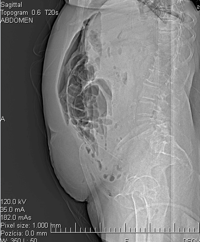
Figure 1: Free air in abdominal cavity.
Immediate laparotomy was performed. The dehiscence of the sutured part of the sigmoid colon and perforation on the lateral side in the aboral part of the sigmoid colon was identified. The surgeon performed Hartmann procedure (left hemicolectomy with sigmoidostomy) with drainage of the pelvic cavity and subhepatic space. After the surgery, the patient was transferred to the intensive care unit, supported by noradrenaline. The antibiotic therapy Ceftriaxone® and Metronidazole® was continued. The next postoperative course was without complications; the drains drain a minimal amount of serosanguinolent content. The drains were extracted on the third postoperative day. The patient was transferred to the department of surgery. The patient's condition gradually improves; the stoma is functional and the laboratory parameters are normal. CRP was 34.92 mg/L on May 14, 2020, when the patient was discharged. The patient with high-grade serous carcinoma, stage IIIC, receives 6 cycles of carboplatin and paclitaxel every 3 weeks.
Discussion
I Incidence, Risks Factors and Pathophysiology
The incidence of intestinal wall injury and its perforation occurs from 0.2% to 1.6% of cases [1, 2]. It can occur during abdominal, vaginal and laparoscopic surgical approaches. It is caused by the unintentional action of mechanical force, thermal energy or their combination on the intestinal wall [2-4]. Several studies have confirmed that the most common damage of the intestine occurs during adhesiolysis and during the entry of instruments into the abdominal cavity during laparoscopic surgery. The lowest incidence is in the case of dilatation and curettage of the uterine cavity [2, 3] . The extent of bowel injury varies and its severity depends on the location of the bowel trauma. During gynaecological procedures, we most often encounter partial damage to the integrity of the visceral peritoneum. Such lacerations of the serous layer of the intestinal wall have the potential to either progress and penetrate the entire thickness of the intestinal wall, or to heal spontaneously during various postoperative periods. The term "enterotomy" or "colotomy" is used in the literature as a synonym for complete intestinal wall penetration. Rarely, the mesentery with vascular structures can be damaged, which causes intestinal tract ischemia [5, 6].
The risk of intestinal wall injury increases with the number of previous laparotomies. Adhesions tend to be present in patients after previous operations in 50% to 80% [1, 3, 6]. In obese patients with body mass index (BMI) of more than 25, the incidence of adhesions is about 90%. There is also an increased risk of intestinal herniation at the scar site, either after laparotomy or laparoscopy. The prevalence of this complication is up to 10% in patients with a BMI of more than 30 [1]. Middle-line laparotomy in combination with the patient's obesity, is a risk factor for the development of umbilical hernia at the scar site, with potential intestinal incarceration.
Small bowel injury may occur during abdominal hysterectomies, but more often, the sigmoid colon is injured due to syntopy of the left ovary and tube. Dissection of pelvic adhesions is a common cause of intestinal injury because bowel loops are fixed into the pelvis by adhesive processes and limited visualization of the pelvic space prevents sufficient visualization and fine adhesiolysis. Adhesive processes are common among patients with endometriosis and deep pelvic inflammation [1, 3, 6]. The patient's age is also one of the risk factors. The patients over 60 years are more often comorbid. Patients with gynaecological malignancies are at risk for iatrogenic bowel injury for various reasons. A history of previous operations and treatment of chemo and radiotherapy leads to the formation of multiple adhesions connecting segments of the intestine with other organs. Repeated laparotomies and extensive lysis of adhesions increase the likelihood of bowel injury. Ovarian malignancies often spread to the intestine, requiring extensive dissection and the resection of the intestine [4, 6, 7]. Patients suffering from endometriosis, deep pelvic inflammation, and after repeated cesarean sections are at a higher risk of bowel injury [2, 6, 7]. Laparotomy is more advantageous for the revision of the abdominal cavity in the case of the suspected intestinal lesions. In the case of laparoscopy, bowel injury is overlooked from 44 to 77% of the patients [5, 8].
The incidence of perforations caused by electro-thermal energy is from 2 to 5 cases per 1000 surgeries and up to 75% are diagnosed more than 7 days after the operation [2, 3, 6]. Thermal damage has a different mechanism, and therefore, the diagnosis and the development of clinical symptoms have a different character than mechanical bowel injury by the surgeon. Thermal tissue damage can be caused by electrosurgical devices through the number of mechanisms, including the unintended direct application of the electrosurgical current, transmission through another conductive instrument, discharge through faulty insulation or capacitive coupling [2]. The extent of intestinal damage caused by bipolar electrocoagulation can be more easily identified than those resulting from monopolar electrocoagulation. If perforation of the entire thickness of the intestinal wall occurs, sufficient excision of the damaged area of the wound margin and its suture is recommended to prevent subsequent suture dehiscence [2, 6, 7]. Thermal damage is often occult, as the visible part of the necrosis is only a small part observed on the surface of the colon. Therefore the presentation of clinical signs may be delayed. Electrocautery is erroneously used as an alternative to simple suture when a defect is located on the serosa of the intestinal wall, increasing the likelihood of late intestinal perforation. However, histological changes in coagulation necrosis are evident only a few hours after ischemic injury, with degenerative changes in cells and their necrosis manifesting within one week, sometimes even after several months [9-11].
During gynaecological operations, we more often encounter mechanical damage of the intestinal wall, especially when adhesions are present. It is usually the result of serous laceration using blunt dissection or cutting directly into the intestinal lumen during a sharp preparation. During adhesiolysis, it is important to apply gentle traction and back pressure to the intestinal loop to facilitate the isolation of the intestine from surrounding tissue and to avoid dissection with sharp scissors or a scalpel. Violent pulling or rough manipulation of the bowel loops can cause the rupture of the bowel wall, resulting in spillage of its contents [4, 5, 11]. Small bowel lesions occur most often during entry into the abdominal cavity and during adhesiolysis. Injuries to the colon usually occur during the pelvic surgery itself. Cytoreductive surgery in ovarian cancer patients may be associated with colonic lesions, even if the surgeon is a certified oncogynaecologist [2, 5, 10].
In laparoscopic procedures, intestinal injury may occur when entering the peritoneal cavity by Veress needle or trocar. The Royal College of Obstetricians and Gynaecologists recommends pressure of 20-25 mmHg pneumoperitoneum and then inserting the trocar with continuous controlled pressure. Before inserting auxiliary trocars, it is important to carefully inspect the abdominal cavity for the presence of adhesion of the omentum and intestines [4, 5, 11, 12]. After laparoscopic as well as laparotomy procedures, herniation of the intestine at the suture site may occur, followed by obstruction and incarceration of the intestine and the development of an ileal condition. Pfannenstiel laparotomy is associated with a lower incidence of herniation than midline laparotomy [1, 6, 12, 13]. Hernias may occur when the fascia is not sutured, namely of 10 mm ports or lager, at the end of the laparoscopy [9, 12, 14]. Callery et al. recommend suturing any fascial defect larger than 5 mm in all layers, including the peritoneum. This reduces the incidence of hernia from 1.83% to 0.17% [9] . Bowel lesions can also occur when the uterine wall is perforated during curettage. An inexperienced surgeon is considered to be a risk factor for this complication [7].
II Clinical Symptoms
Intestinal perforation should always be considered in the differential diagnosis of a worsened postoperative course. Typical symptoms may appear already a few hours after surgery. A patient with intestinal injury presents clinically with vagal symptoms: nausea and vomiting. There is a colic abdominal pain with palpation sensitivity in the whole abdomen and the overall discomfort of the patient. The clinical picture resembles postoperative ileus, accompanied by peritoneal irritation, fever and leukocytosis. Significant abdominal distension, meteorism and palpation diffuse sensitivity are present as a manifestation of diffuse peritonitis [1, 2, 11]. Colon injuries can have much more serious consequences and a faster alteration of patient‘s health status. The threat of complete perforation of the colon is the spillage of feces into the abdominal cavity, leading to stercoral peritonitis. Bacterial inflammation is accompanied by an elevation of inflammatory markers, leukocytes, C-reactive protein and procalcitonin. If an intestinal injury is unrecognized, there is an alteration of the patient's mental status with tachycardia, tachypnoea, and febrility as the surgical response to an infectious stimulus, which can progress to septic shock and multiorgan failure syndrome.
III Diagnosis
Early detection of intestinal damage and early intervention are crucial for reducing morbidity and mortality. Despite rigorous inspection, only half of the cases are found to have bowel damage during surgery. If bowel damage is not diagnosed and treated during primary surgery, it can become a life-threatening condition for the patient. Elbiss and Abu-Zidan emphasize that the mortality rate increases significantly if the diagnosis of the intestinal injury is delayed by more than three days [11]. The symptoms of bowel damage manifest obviously within 12-36 hours after surgery, but they can appear 5 or 7 days later. This can happen in the case of delayed necrosis with intestine thermal damage, or the development of a pericolic abscess, which perforates later after the surgery. Patients may develop non-specific symptoms such as abdominal pain, oral intolerance, bloating, nausea, febrile, diarrhea, which may delay diagnosis. Late presentation of symptoms includes generalized peritonitis, abscess formation, and septic shock. Repeated abdominal examinations performed every 4-6 hours are important for proper diagnosis and subsequent surgery [4, 7, 10, 11, 15] .
During dilation, curettage, hysteroscopic and laparoscopic surgical procedure, the intestinal injury might not be immediately recognized and it is necessary to monitor the development of associated clinical symptoms of peritoneal irritation, which may indicate either a haemorrhage or perforation of the intestine [13, 15]. However, an examination of the abdomen in severe sepsis, associated with alterations in the mental status of ventilated patients, can be misleading. It is important to monitor inflammatory parameters, such as leukocyte count, procalcitonin and C-reactive protein, which are very sensitive to changes in the internal environment and can thus significantly help in diagnosis. Continuous rising of procalcitonin and C-reactive protein is significant for bacterial inflammation caused by fecal spillage of the abdominal cavity. They are also very useful in the postoperative monitoring of the patient, because their values decrease rapidly after the insult has resolved [2].
The diagnosis can be determined by several auxiliary examinations. X-ray of the abdomen verifies the free air in the abdominal cavity. Ultrasound of the abdomen describes the presence of free air in the abdominal cavity as increased echogenicity compared to the echogenicity of intra-abdominal organs. However, contrast computed tomography (CT) is the gold standard in the diagnosis of intestinal perforation. It describes not only the presence of free air in the abdominal cavity but also the presence of fluid collection in the form of a pericolic abscess. CT of the abdomen should be performed in every case of any suspicion of intestinal injury. Monitoring of renal and liver function as well as electrolyte fluid balance should be performed in such cases. Delayed diagnosis of bowel perforation seriously threatens patient's life and very often initializes the forensic consequences [1, 2, 11, 15].
IV Treatment
The treatment of an intestinal injury depends on the location of the lesion and its extent. If the gynaecologist is experienced in bowel surgery, simple lacerations and small perforations, then the patient can be treated without attending a surgeon. Due to possible forensic consequences, the preferred way is calling for the surgeon. Serosal tears represent weak points in the bowel. If bowel obstruction and subsequent distension develop postoperatively, these weak spots may perforate, leading to peritonitis or enterocutaneous fistulae. Therefore, they should be treated carefully [3, 7]. When the injury is less than half of the circumference of the injured segment of the bowel, a repair can be performed without bowel resection. The suture of the defect must be in a perpendicular axis to the course of the intestine to prevent narrowing of the lumen. The bowel wall can be sutured in one or two layers [3, 7]. When a single-layer closure is chosen, Mendez prefers to use the Gambee technique that has been shown to be safe and efficacious [5].
When the injury is longer than half of the circumference of the bowel or when the simple repair causes a stricture of the lumen to less than 1 to 2 cm in diameter, resection and end-to-end anastomosis are indicated. It is more appropriate to perform a temporary ileostomy or jejunostomy to prevent suture dehiscence on a patient after radiotherapy and when the perforation site is in the irradiated area [5]. The large bowel has a larger lumen than does the small bowel, and therefore does not require closing the laceration perpendicularly to the intestinal lumen in an attempt to avoid stricturing and subsequent obstruction. Suture of a section of intestine longer than 2 cm should be sutured in two layers, similar to the small intestine. Resection and anastomosis are indicated when the laceration is greater than 30%-40% of the circumference of the injured segment. A colostomy is the safest approach to treat large bowel injuries due to the increased incidence of complications [5, 15].
Forensic Consequences
Bowel injury during surgery from serosal tearing to complete bowel perforation is a serious complication. Subsequent colostomy affects the patient's quality of life. The best prevention includes careful analysis of the history of the patient, the precise planning of the type of surgical procedure and surgeon experience and certification in bowel surgery. The consent form should contain information about potential complications during surgical procedure and should be written by the patient and the surgeon [16]. The communication with the patient and her relatives is critical when such complications occur. Documentation with the precise description of the diagnostic and treatment methods is imperative in the prevention of the forensic consequences. Regular morbidity and mortality meeting have the aims to educate the staff as well as the use of the guidelines to minimize these cases [16].
Acknowledgements
None.
Conflicts of Interest
None.
Consent
The written consent for publication was taken from the patient.
Article Info
Article Type
Case Report and Review of the LiteraturePublication history
Received: Mon 20, Jul 2020Accepted: Mon 03, Aug 2020
Published: Tue 11, Aug 2020
Copyright
© 2023 Miloš Mlynček. This is an open-access article distributed under the terms of the Creative Commons Attribution License, which permits unrestricted use, distribution, and reproduction in any medium, provided the original author and source are credited. Hosting by Science Repository.DOI: 10.31487/j.SCR.2020.08.12
Figures & Tables
References
- Roger Kirby, Frank Arnold, Ben Challacombe, Krishna Patil, Peter Amoroso et al. (2011) Diagnosis and management of bowel injury during laparoscopic surgery. Trends Urol Men´s Health 18-21.
- Sebastiano Cassaro (2015) Delayed Manifestations of Laparoscopic Bowel Injury. Am Surg 81: 478-482. [Crossref]
- Daniel Paloyan (2008) Intestinal Problems in Gynecologic Surgery. Glob Libr Women´s Med 10: 1-11.
- Elaheh Mesdaghinia, Masoumeh Abedzadeh Kalahroudi, Mehrdad Hedayati, Nushin Moussavi Bioki (2013) Iatrogenic Gastrointestinal Injuries During Obstetrical and Gynecological Operation. Arch Trauma Res 2: 81-84. [Crossref]
- L E Mendez (2001) Iatrogenic Injuries in Gynecologic Cancer Surgery. Surg Clin North Am 84: 897-923. [Crossref]
- Meng TzungWu, Lim WohKoh, Song Nan Chow (2004) Can Bowel Injury be Prevented During Laparoscopic Surgery? A Case Report and Literature Review. Taiwan J Obstet Gynecol 43: 219-221.
- James D Perkins, leon L Dent (2004) Avoiding and Repairing Bowel Injury in Gynecologic Surgery. OBG Manag 16: 15-28.
- Cheng Xiang Shan, Chong Ni, Ming Qiu, Dao Zhen Jiang (2012) Is Laparoscopy Equal to Laparotomy in Detecting and Treating Small Bowel Injuries in a Porcine Model? World J Gastroenterol 18: 6850-6855. [Crossref]
- Kerstin Bewö, Johanna Österberg, Mats Löfgren, Gabriel Sandblom (2019) Incisional Hernias Following Open Gynecological Surgery: a Population-Based Study. Arch Gynecol Obstet 299: 1313-1319. [Crossref]
- C Chapron, F Pierre, Y Harchaoui, S Lacroix, S Béguin S et al. (1999) Gastrointestinal Injuries during Gynaecological Laparoscopy. Hum Reprod 14: 333-337. [Crossref]
- Hassan M Elbiss, Fikri M Abu Zidan (2017) Bowel Injury Following Gynecological Laparoscopic Surgery. Afr Health Sci 17: 1237-1245. [Crossref]
- Marilyn Huang, Fernanda Musa, Caroline Castillo, Kevin Holcomb (2010) Postoperative Bowel Herniation in a 5-mm Nonbladed Trocar Site. JSLS 14: 289-291. [Crossref]
- C V Hegde (2012) Management of Large Bowel Injury during Laparoscopic Surgery. J Obstet Gynaecol India 62: 501-503. [Crossref]
- Hitoshi Tonouchi, Yukinari Ohmori, Minako Kobayashi, Masato Kusunoki (2004) Trocar Site Hernia: A more Common Problem than We Believe? Arch Surg 139: 1248-1256. [Crossref]
- Kahraman Ulker, Turgut Anuk, Murat Bozkurt, Yetkin Karasu (2014) Large Bowel Injuries during Gynecological Laparoscopy. World J Clin Cases 2: 846-851. [Crossref]
- Paul A Walden, Burak Zeybek, John Y Phelps (2018) Understanding the Legal Essentials of a Bowel Injury Lawsuit in Minimally Invasive Gynecologic Surgery. J Minim Invasive Gynecol 25: 30-37. [Crossref]