Surgical Ciliated Cyst of the Maxilla: A Case-Series of Three Cases
A B S T R A C T
The surgical ciliated cyst is an iatrogenic lesion occurring after surgeries in which the Schneiderian membrane has been exposed, such as in orthognathic surgery or maxillary sinus procedures. This lesion has been infrequently documented in western countries. In this case series, we present three cases of surgical ciliated cysts of the maxilla.
Keywords
Surgical ciliated cyst, sinus, pathology
Introduction
Surgical ciliated cysts, also called post-operative maxillary cysts, are benign, frequently iatrogenic cysts of the jaws that develop after maxillofacial surgical procedures. These cysts develop after inadvertent implantation of the epithelial lining of the maxillary sinus into adjacent maxillary bone following procedures such as the Le Fort 1 osteotomy, Caldwell-Luc antrostomy, and complex surgical extraction of posterior maxillary teeth [1]. Surgical ciliated cysts are characterized radiographically by well demarcated, unilocular radiolucencies of varying sizes located in the surrounding bone but separate from the sinus [2]. Additionally, there have been reports of rare but well-documented surgical ciliated cysts being found in the mandible, presumably caused by surgical instruments contaminated with sinus membrane epithelium used in orthognathic procedures involving maxillary and mandibular bones simultaneously [3]. The surgical ciliated cyst is a rare form of pathology, and there have been only 10 case reports from western countries in the literature over the last 15 years. However, these cystic lesions are more common in Japan, which was hypothesized to be due to either ethnic differences, misdiagnosis, or underdiagnosis [1, 4]. Surgical curettage is the mainstay for the treatment of surgical ciliated cysts. The intent of this article is to present three additional cases diagnosed at the Dental College of Georgia at Augusta University of this relatively rare iatrogenic lesion and review the relevant literature.
Case Reports
Case Report I
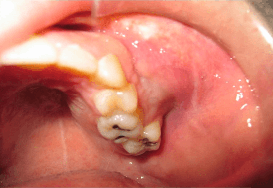
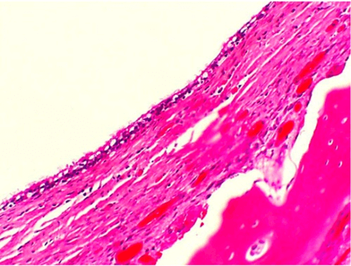
Case I is a 26-year-old Caucasian male who received an open reduction and internal fixation of a Lefort I fracture after an automobile accident. The patient subsequently reported to the Oral and Maxillofacial Surgery Clinic at the Dental College of Georgia six months post-operatively with complaints of pain and swelling in the left mid-face region. Clinical examination revealed slight fullness in the left mid-face region. Upon intraoral examination, there was an expansion of both buccal and palatal cortical plates in the left maxillary premolar/molar region with mobility of teeth 12, 14, and 15 (Figure 1). The panoramic radiograph depicts a well-demarcated radiolucency associated with the fixation plate and screws on the left zygomatic buttress (Figure 2). Resorption of the maxillary posterior teeth roots was also noted. Total enucleation of the lesion was performed with removal of the adjacent maxillary hardware and submission of the specimen for pathologic report (Figures 3 & 4). This case was signed out as an inflamed surgical ciliated cyst.
Figure 1: Expansion of buccal and lingual cortical plates.
Figure 2: Arrow depicting well-demarcated radiolucency associated with surgery site.
Figure 3: Fixation screws floating in the lesion in left maxilla.
Figure 4: Histologic examination with HE staining revealed a soft tissue specimen consisting of multiple sections of a moderately cellular fibrous capsule lined by ciliated columnar epithelium with peripheral layers of bone, adherent periosteum, and muscle.
Case Report II
Case II is of a 39-year-old Caucasian female who received a Lefort I osteotomy to correct a maxillary deficiency. The patient reported 1-year post-operatively complaining of pain and tenderness of both cheeks. Radiographs reveal bilateral radiolucencies associated with fixation plates and screws placed during the previous orthognathic surgery (Figure 5). Treatment was total enucleation of the lesion with the removal of the associated hardware and submission of the specimen for pathologic report (Figure 6). The case was signed out as a surgical ciliated cyst.
Figure 5: Arrows depicting well-demarcated bilateral radiolucencies associated with hardware from Lefort I osteotomy.
Figure 6: Histologic examination with HE staining revealed a densely collagenized fibrous connective tissue capsule lined by respiratory epithelium. The fibrous capsule is thickened and supports hemorrhagic foci and aggregates lymphocytes, histiocytes, and hemosiderin laden macrophages.
Case Report III
A 53-year-old Hispanic female presented with a 10mm radiopaque solitary unilocular lesion in the left maxillary sinus present for about 5 years (Figure 7). The patient had an unremarkable medical history but did have a left Caldwell-Luc sinus lift in 2014. Curettage of the lesion was performed, revealing a well-defined bony cavity with communication to the sinus membrane (Figure 8). The case was signed out as a surgical ciliated cyst (Figure 9).
Figure 7: Panoramic radiograph showing a 10mm radiopaque solitary unilocular lesion in the left maxillary sinus.
Figure 8: Surgical exposure of the cyst lining in the left maxillary sinus.
Figure 9: Histologic examination revealed a soft tissue specimen consisting of a fibrous capsule lined by respiratory epithelium.
Discussion
It is widely understood that the development of surgical ciliated cysts is due to the entrapment of sinus mucosa in the bone surrounding the wound created during orthognathic surgeries, maxillary sinus surgeries, or other procedures involving exposure of the Schneiderian membrane [1]. Although they normally occur in maxillary bone, there have been some reports of surgical ciliated cyst in the mandible as well [3]. Radiographically, they appear as well defined, unilocular radiolucencies associated with, but separate from, the maxillary sinus. Histologically, they are characterized by moderately cellular fibrous capsules lined by ciliated columnar epithelium consistent with the Schneiderian membrane. Despite a low number of surgical ciliated cysts reported in the literature from the western hemisphere, the surgical ciliated cyst should be considered in the differential diagnosis of cystic lesions occurring after surgical procedures in which the maxillary sinus lining has been breached, such as those listed above [4-14].
Cases of surgical ciliated cysts can present with or without symptoms. Signs and symptoms include pain, tenderness, and swelling of the midface region. The mainstay treatment for surgical ciliated cysts is enucleation and curettage. If the size of a surgical ciliated cyst increases to the point of damaging surrounding structures, marsupialization is indicated. Recurrences of surgical ciliated cysts are not well documented in the literature. Further research is required to explain the low number of cases of surgical ciliated cysts diagnosed in the western hemisphere and the recurrence rate of this pathologic entity.
Conflicts of Interest
None.
Article Info
Article Type
Case SeriesPublication history
Received: Mon 29, Nov 2021Accepted: Sat 18, Dec 2021
Published: Wed 29, Dec 2021
Copyright
© 2023 Andrew Jenzer. This is an open-access article distributed under the terms of the Creative Commons Attribution License, which permits unrestricted use, distribution, and reproduction in any medium, provided the original author and source are credited. Hosting by Science Repository.DOI: 10.31487/j.DOBCR.2021.04.01
Author Info
Macarius Abdelsayed Jeffrey James Kyle B Frazier Brian Sellers Rafik Abdelsayed Andrew Jenzer
Corresponding Author
Andrew JenzerStaff Surgeon, Residency, Department of Oral and Maxillofacial Surgery, Eisenhower Army Medical Center, Augusta, Georgia, USA
Figures & Tables


References
1. Golaszewski J,
Muñoz R, Barazarte D, Perez L (2019) Surgical ciliated cyst after maxillary
orthognathic surgery: a literature review and case report. Oral Maxillofac
Surg 23: 281-284. [Crossref]
2. Yamamoto S, Maeda
K, Kouchi I, Hirai Y, Taniike N et al. (2017) Surgical Ciliated Cyst Following
Maxillary Sinus Floor Augmentation: A Case Report. J Oral Implantol 43:
360-364. [Crossref]
3. Seifi S, Sohanian
S, Khakbaz O, Abesi F, Aliakbarpour F et al. (2016) Ectopic Ciliated Cyst in
the Mandible Secondary to Genioplasty and Lefort after Two Years: A Case Report
and Literature Review. Iran J Otorhinolaryngol 28: 353-356. [Crossref]
4. Leung YY, Wong WY,
Cheung LK (2012) Surgical Ciliated Cysts May Mimic Radicular Cysts or Residual
Cysts of Maxilla: Report of 3 Cases. J Oral Maxillofac Surg 70:
e264-e269. [Crossref]
5. Li CC, Feinerman
DM, MacCarthy KD, Woo SB (2014) Rare mandibular surgical ciliated cysts: report
of two new cases. J Oral Maxillofac Surg 72: 1736-1743. [Crossref]
6. Sugar AW, Walker
DM, Bounds GA (1990) Surgical ciliated (postoperative maxillary) cysts
following mid-face osteotomies. Br J Oral Maxillofac Surg 28: 264-267. [Crossref]
7. Hayhurst DL,
Moenning JE, Summerlin DJ, Bussard DA (1993) Surgical ciliated cyst: a delayed
complication in a case of maxillary orthognathic surgery. J Oral Maxillofac
Surg 51: 705-709. [Crossref]
8. Niederquell BM,
Brennan PA, Dau M, Moergel M, Frerich B et al. (2016) Bilateral Postoperative
Cyst after Maxillary Sinus Surgery: Report of a Case and Systematic Review of
the Literature. Case Rep Dent 2016: 6263248. [Crossref]
9. Kim JJ, Freire M,
Yoon JH, Kim HK (2013) Postoperative maxillary cyst after maxillary sinus
augmentation. J Craniofac Surg 24: e521-e523. [Crossref]
10. Shakib K, McCarthy
E, Walker DM, Newman L (2009) Post operative maxillary cyst: report of an
unusual presentation. Br J Oral Maxillofac Surg 47: 419-421. [Crossref]
11. Basu MK, Rout PG,
Rippin JW, Smith AJ (1988) The post-operative maxillary cyst. Experience with
23 cases. Int J Oral Maxillofac Surg 17: 282-284. [Crossref]
12. Amin M, Witherow H,
Lee R, Blenkinsopp P (2003) Surgical ciliated cyst after maxillary orthognathic
surgery: report of a case. J Oral Maxillofac Surg 61: 138-141. [Crossref]
13. Pe MB, Sano K, Kitamura A, Inokuchi T (1990) Computed tomography in the evaluation of postoperative maxillary cysts. J Oral Maxillofac Surg 48: 679-684. [Crossref]
14. Bulut AŞ, Sehlaver C, Perçin AK (2010) Postoperative maxillary cyst: a case report. Patholog Res Int 2010: 810835. [Crossref]
