Journals
The Value of Longitudinal Strain versus Coronary Angiography in Detection of Coronary Artery Disease
A B S T R A C T
Aims: The aim of this study was to evaluate the value and accuracy of longitudinal strain in detection of coronary artery disease compared to coronary angiography. Results: The left ventricular longitudinal strain-speckle tracking showed evidence of stenosis of left anterior descending artery, circumflex artery and right coronary artery in (86.1%), (76.4%), and (84.7%) respectively. For the stenosis in left anterior descending artery, the current study showed that the longitudinal strain was a good predictor for presence of significant stenosis with a sensitivity of (93.8%), specificity (75%) and accuracy (91.7%) compared with coronary angiography. For the stenosis in right coronary artery, the left ventricular longitudinal strain had a sensitivity of (93.5%), specificity of (70.0%) and accuracy of (90.3%) compared with coronary angiography. Conclusion: Speckle-tracking echocardiography technique has a good performance and validity in detection of coronary artery stenosis with good agreement with angiography.
Keywords
longitudinal strain, speckle tracking, coronary artery disease
I N T R O D U C T I O N
Coronary artery disease is characterized by atherosclerosis in the epicardial coronary arteries. The reduction in coronary artery flow may be symptomatic or asymptomatic, occurs with exertion or at rest, and culminate in myocardial infarction or angina, depending on obstruction severity and the rapidity of development [1].
Recent studies have suggested that decreased left ventricular (LV) compliance accompanies both coronary artery disease and acute myocardial infarction. Many facilities measure only left ventricular ejection fraction (LVEF) and left ventricular end-diastolic pressure (LVEDP) when assessing left ventricular dysfunction in patients with coronary artery disease (CAD) [2, 3]. Speckle-tracking echocardiography is a new noninvasive ultrasound imaging technique that allows for an objective and quantitative evaluation of global and regional myocardial function independently from the angle of insonation and from cardiac translational movements [4-7].
Patients and Methods
Study Design
A cross sectional study was conducted in Ibn Al-Bittar Cardiac Surgical Center/ Baghdad. It included a sample of 72 patients with coronary artery disease proved by coronary angiography and then echocardiography for each patient was done. Also, evaluation of risk factors of CAD like smoking, diabetes mellitus, hypertension, lipid profile along with body mass index were taken in consideration.
The study was conducted in compliance with the medical ethics rules and all participants have given their consent. The study protocol was approved by the ethical committee of the department of medicine/ Baghdad medical school.
Types of investigations used
All the study subjects were evaluated by treadmill test TMT (which is considered positive TMT if there is ST segment deviation one mm or more in one or more ECG leads and/or if the patient develops ischemic chest pain). Lipid profile, conventional echocardiography & 2D LV Longitudinal strain using speckle tracking analysis by the available 2D strain software (GE Vivid E9) within 1wk of the scheduled coronary angiography.
Conventional echocardiography
Conventional echocardiography was done using echocardiography machine (GE Vivid E-9) and phased-array probe with a frequency of 2-4 MHz.
Evaluation of LV systolic function (LV ejection fraction) was done using linear M-mode echocardiography with M-mode cursor at the tips of mitral valve leaflets in parasternal long axis view. Regional wall motion abnormality was assessed using apical long axis view for assessment of anterio-septal and posterior-lateral walls at basal, mid, apical segments; LV 4-chamber view for septal and lateral walls at basal, mid, apical segments; LV 2-chamber view for anterior and inferior walls at basal mid apical segments, correlated with LV short axis view at basal, mid, and apical levels.
Two-dimensional speckle tracking analysis
Using available 2D strain software (GE E-9), acquisition of 3 apical views (2-chamber, 3-chamber and 4- chamber) was done. All images were taken at end expiration to avoid LV apical foreshortening and reducing sector width and depth was used to increase frame rate for better image resolution. All images were digitally stored at a high frame rate (>63 frames/s, mean 70 ± 12).
Region of interest was localized at LV endocardial border 2 at base, and one apical to include the entire myocardium at end systolic frame. The software algorithm automatically segmented the LV into six equidistant segments and selected suitable speckles in the myocardium for tracking. The software algorithm then tracked the speckle patterns on a frame-by-frame basis using the sum of absolute difference algorithm. Finally, the software automatically generated LV strain profiles for each of the six segments of each view, from which end-systolic strain was measured. The average value of strain at each level (basal, middle, and apical) and global strain obtained using Bull eye model measuring peak regional & global LV longitudinal strain of 17 LV segments was calculated.
All measurements were preformed off-line on a dedicated workstation using available 2D strain software (GE E9). The examiner was blinded to patient's results of coronary angiography. Longitudinal strain was computed using 2D speckle-tracking analysis by automated function imaging (AFI). For strain processing, the peak of the R-wave on the electrocardiogram was used as the reference time point for end-diastole and segments with poor-quality tracking were manually discarded. GLS was only computed from patients with >92% of segments adequately tracked (≥15 segments for a 17-segment model). Given the average value of (–17 ± 0.1%) for normal GLS, any value assessed by AFI less than (−16%) was considered as impaired GLS.
Thorough correlation of LV regional longitudinal impairment with coronary territories was evaluated in all patients, value of regional longitudinal strain less than (−16%) were abnormal and diagnostic of CAD if it involved more than 3 segments in same coronary territories.
Coronary angiography
Coronary angiography was done for all patient using standard protocol and the diagnosis of CAD was defined if there is stenosis more than or equal to 50% in left main coronary artery (LMS) and/ or if the stenosis was more than or equal to 70% in the right coronary artery (RCA), left anterior descending coronary artery (LAD) and/ or circumflex coronary artery (CX).
Statistical analysis
Data of the patients were entered and analyzed using the statistical package for social sciences (SPSS) version 22, IBM Inc., Chicago, USA, 2013. Descriptive statistics were presented as means, standard deviations, ranges, frequencies and proportions according to the variable types. Cross-tabulation and Receiver operating characteristics (ROC) curve analysis was used to assess the validity of the 2D LV Longitudinal strain -speckle tracking in detection of stenosis in different coronary arteries, kappa statistics used to assess the performance and the percent agreement of the 2D LV Longitudinal strain -speckle tracking in detection in comparison with angiography. Level of significance was set at 0.05 to be significant difference or correlation. Finally results and findings were presented in tables, figures and explanatory paragraphs using Microsoft office (Word), 2013 software for windows.
Results
The baseline demographic characteristics of the patients are shown in (Table 1). Regarding age, the mean age of patients was (56.4 ± 10.1 yrs), and the mean body mass index (BMI) was (27.2 ± 3.2 kg/m2). Only 20 patients (27.8%) had BMI within normal levels (BMI < 25 kg/m2). The smokers represented about half of the patients (51.4%), 53 patients (73.6%) were diabetic, 24 (33.3%) were hypertensive, 29 (40.3%) had pericardial fat and 42 patients (58.3%) had hyperlipidemia.
Table 1: Demographic characteristics of the study population
|
Characteristic |
|
N= 72 |
|
|
Age (year) |
Mean± SD |
56.4 ± 10.1 |
|
|
BMI category |
Mean± SD |
27.2 ± 3.2 |
|
|
BMI
|
(Normal) |
|
20 (27.8%) |
|
(Overweight) |
|
35 (48.6%) |
|
|
(Obese) |
|
17 (23.6%) |
|
|
Smoking |
(Yes) |
|
37 (51.4%) |
|
(No) |
|
35 (48.6%) |
|
|
Diabetes mellitus |
(Yes) |
|
53 (73.6%) |
|
(No) |
|
19 (26.4%) |
|
|
Hypertension |
(Yes) |
|
24 (33.3%) |
|
(No) |
|
48 (66.7%) |
|
|
Pericardial Fat |
(Yes) |
|
29 (40.3%) |
|
(No) |
|
43 (59.7%) |
|
|
Hyperlipidemia |
(Yes) |
|
42 (58.3%) |
|
(No) |
|
30 (41.7%) |
|
According to the angiographic findings, it had been found that left anterior descending artery (LAD) stenosis was present in 64 patients (88.9%), the circumflex coronary artery (LCX artery) stenosis in 56 patients (77.8%), and the right coronary artery (RCA) stenosis in 62 patients (86.1%) (Table 2).
According to the findings of 2-D LV longitudinal strain-speckle tracking, positive finding of left anterior descending artery (LADA) stenosis was present in 62 patients (86.1%), the circumflex (CX) artery stenosis was found in 55 patients (76.4%) and the right coronary artery (RCA) stenosis was found in 61 patients (84.7%) (Table 3).
Table 2: Angiographic findings of the study group (N = 72)
|
|
Stenosis |
|||
|
Coronary artery |
Present |
Absent |
||
|
|
No |
% |
No |
% |
|
Left anterior descending |
64 |
88.9 |
8 |
11.1 |
|
Circumflex |
56 |
77.8 |
16 |
22.2 |
|
Right |
62 |
86.1 |
10 |
13.9 |
Table 3: Findings of 2-D LV Longitudinal strain –speckle tracking in study patients
|
|
Stenosis |
|||
|
Coronary artery |
Present |
Absent |
||
|
|
No |
% |
No |
% |
|
Left anterior descending |
62 |
86.1 |
10 |
13.9 |
|
Circumflex |
55 |
76.4 |
17 |
23.6 |
|
Right |
61 |
84.7 |
11 |
15.3 |
Table 4: Shows the number of stenosed arteries detected by both methods.
|
Method |
Number of diseased arteries |
No. |
% |
|
Angiography |
One vessel |
9 |
12.5 |
|
Two vessels |
16 |
22.2 |
|
|
Three vessels |
47 |
65.3 |
|
|
2D LV Longitudinal strain |
One vessel |
10 |
13.9 |
|
Two vessels |
18 |
25.0 |
|
|
Three vessels |
44 |
61.1 |
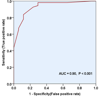
Table 5: shows the cross-tabulation of the findings of longitudinal strain against angiography regarding the stenosis in left anterior descending artery (LADA); this revealed that longitudinal strain was a good predictor for LADA stenosis with a sensitivity of (93.8%), specificity of (75%) and accuracy of (91.7%). On the other hand, kappa value was 0.82 which indicated good performance of the test and a percent agreement of 91.8% between both tests (P-value < 0.001). Furthermore, ROC curve revealed an area under the curve (AUC) of 0.90 which indicated good predictive value and accuracy of the test (P < 0.001) (Fig. 1).
Table 5: Validity and performance of 2-D LV longitudinal strain-speckle tracking in detection of left anterior descending artery stenosis.
|
Longitudinal strain |
Angiography |
Total |
||||
|
Positive |
Negative |
|||||
|
No. |
% |
No. |
% |
No. |
% |
|
|
Positive |
60 |
93.8 |
2 |
25.0 |
62 |
86.1 |
|
Negative |
4 |
6.3 |
6 |
75.0 |
10 |
13.9 |
|
Total |
64 |
100.0 |
8 |
100.0 |
72 |
100.0 |
|
Validity test |
||||||
|
Sensitivity 93.8% |
||||||
|
Specificity 75.0% |
||||||
|
Accuracy 91.7% |
||||||
|
PPV (positive predictive value) 96.8% |
||||||
|
NPV (negative predictive value) 60.0% |
||||||
|
Kappa = 0.82, Percent agreement = 91.8%, P-value < 0.001 |
||||||

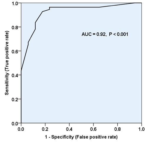
In (table 6), the longitudinal strain showed good performance in detection of stenosis in LCX artery with a sensitivity of (92.9%), specificity of (81.3%) and accuracy of (90.3%), (kappa = 0.82, percent agreement = 90.2%, P< 0.001), (Fig. 2).
Table 6: Validity and performance of 2-D LV Longitudinal strain-speckle tracking in detection of left circumflex coronary artery stenosis.
|
Longitudinal strain |
Angiography |
Total |
||||
|
Positive |
Negative |
|||||
|
No. |
% |
No. |
% |
No. |
% |
|
|
Positive |
52 |
92.9 |
3 |
18.8 |
55 |
76.4 |
|
Negative |
4 |
7.1 |
13 |
81.3 |
17 |
23.6 |
|
Total |
56 |
100.0 |
16 |
100.0 |
72 |
100.0 |
|
Validity test |
||||||
|
Sensitivity 92.9% |
||||||
|
Specificity 81.3% |
||||||
|
Accuracy 90.3% |
||||||
|
PPV (positive predictive value) 94.5% |
||||||
|
NPV (negative predictive value) 76.5% |
||||||
|
Kappa = 0.82, Percent agreement = 90.2%, P-value < 0.001 |
||||||

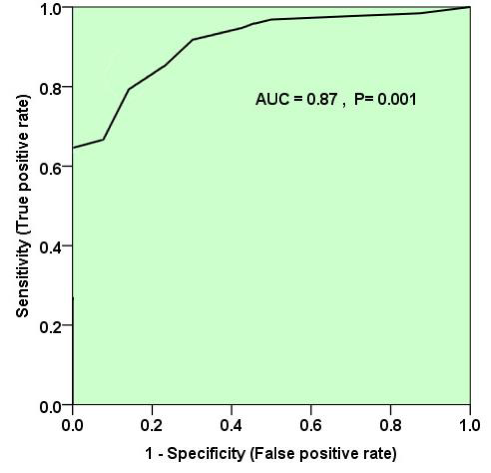
Regarding the performance and validity of 2-D LV longitudinal strain in detection of RCA stenosis in comparison with angiography, it had a sensitivity of (93.5%), specificity of (70.0%) and accuracy of (90.3%), (kappa = 0.88, percent agreement = 90.2%, P< 0.001), (Table 7), (Fig. 3).
Table 7: Validity and performance of 2D LV Longitudinal strain – speckle tracking in detection of RCA stenosis.
|
Longitudinal strain |
Angiography |
Total |
||||
|
Positive |
Negative |
|||||
|
No. |
% |
No. |
% |
No. |
% |
|
|
Positive |
58 |
93.5 |
3 |
30.0 |
61 |
84.7 |
|
Negative |
4 |
6.5 |
7 |
70.0 |
11 |
15.3 |
|
Total |
62 |
100.0 |
10 |
100.0 |
72 |
100.0 |
|
Validity test |
||||||
|
Sensitivity 93.5% |
||||||
|
Specificity 70.0% |
||||||
|
Accuracy 90.3% |
||||||
|
PPV (positive predictive value) 95.1% |
||||||
|
NPV (negative predictive value) 63.6% |
||||||
|
Kappa = 0.88, Percent agreement = 90.2% , P-value < 0.001 |
||||||

Additionally, the validity of longitudinal strain in detection of stenosis in different numbers of coronary arteries are shown in (Table 8), (Kappa = 0.86, Percent agreement = 84.7%, P-value < 0.001), hence, the validity and performance in detection of multiple vessel stenosis were good and the best values reported in detection of three-vessels disease.
Table 8: Validity and performance of 2-D LV Longitudinal strain-speckle tracking in detection of different numbers of stenosed coronary arteries
|
|
Angiography |
Total |
||||||
|
Longitudinal strain |
One vessel |
Two vessels |
Three vessels |
|||||
|
No. |
% |
No. |
% |
No. |
% |
No. |
% |
|
|
One vessel |
7 |
77.8 |
3 |
18.8 |
0 |
0.0 |
10 |
13.9 |
|
Two vessels |
2 |
22.2 |
11 |
68.8 |
4 |
8.5 |
17 |
23.6 |
|
Three vessels |
0 |
0.0 |
2 |
12.5 |
43 |
91.5 |
45 |
62.5 |
|
Total |
9 |
100 |
16 |
100 |
47 |
100 |
72 |
100 |
|
Validity test |
One vessel |
|
Two vessels |
|
Three vessels |
|
|
|
|
Sensitivity 77.8% 68.8% 91.5% |
||||||||
|
Specificity 95.2% 88.5% 92.0% |
||||||||
|
Accuracy 93.1% 79.2% 91.7% |
||||||||
|
PPV(positive predictive value) 70.0% 64.7% 95.6% |
||||||||
|
NPV (negative predictive value) 96.8% 83.6% 85.2% |
||||||||
|
Kappa = 0.86, Percent agreement = 84.7% , P-value < 0.001 |
||||||||
Discussion
Speckle Tracking Echocardiography (STE) is an echocardiographic imaging technique that analyzes the motion of tissues in the heart by using the naturally occurring speckle pattern in the myocardium when imaged by ultrasound. This novel method of documentation of myocardial motion represents a noninvasive method of definition of vectors and velocity. When compared to other technologies seeking noninvasive definition of ischemia, speckle tracking seems a valuable endeavor. This speckle pattern is a mixture of interference patterns and natural acoustic reflections [8]. These reflections are also described as speckles or markers. The pattern is being random, each region of the myocardium, has a unique speckle pattern (also called patterns, features, or fingerprints), that allows the region to be traced from one frame to the next, and this speckle pattern is relatively stable, at least from one frame to next [9, 10].
The current study shows that the main age group of patients was between 50-59 years with mean age 56.4 ± 10.1 years, which is the same that was found by Cimino et al., and by Montgomery et al [11, 12].
Smoking is a major risk factor for coronary heart disease which is also found in the present study that shows that more than half of the studied group were smokers; this is consistent with that found by Cimino et al., where about 59% of patients were smokers [11].
In asymptomatic diabetic patients with a preserved LVEF, it has been shown that speckle-tracking echocardiography has the potential for detecting subclinical LV systolic dysfunction, which is unmasked by the alteration of longitudinal strain. In this view, speckle-tracking echocardiography might provide useful information about the development of subclinical myocardial dysfunction in the diabetic setting before the overt appearance of diabetic cardiomyopathy. This evidence confirms previous experiences using either color tissue velocity imaging or doppler-derived strain rate imaging [13]. The current study shows that about two thirds of the participants had diabetes mellitus which is the same finding that was reported by Tsutomu Takagi [14].
The present study shows that about one third of the patients were suffering from hypertension, which in accordance with that revealed by Ji Qiang et al [15].
Regarding the hyperlipidemia, it was found that more than half of the patients had hyperlipidemia, which is less than found by Cimino et al. where more than two thirds of the patients suffer from it. This may be due to that the patients in the present study were diagnosed previously and were on treatment which includes cholesterol lowering medications [11].
For the body mass index (BMI), the current study shows that about half of the patients were overweight and the mean BMI was (27.2 kg/m2) which is concordant with that revealed by Lipinski, Michael et al., where it was (28.5%) [16].
According to the angiographic findings, it was found that left anterior descending coronary artery (LAD) stenosis was reported in (88.9%), the left circumflex coronary artery (LCX) stenosis reported in (77.8%) and the right coronary artery (RCA) stenosis was found in (86.1%) of the patients while in a study done in USA by Montgomery, David et al 12 the results were: the stenosis in LAD only (21%), in LCX only (9%) and the stenosis in RCA only (7%). Additionally, it was (25%) in stenosis of LAD + LCX, (11%) in LAD + RCA and it was (4%) for the stenosis in LCX + RCA.
In the present study, 2-D LV Longitudinal strain-speckle tracking showed evidence of stenosis of LAD (left anterior descending artery) in (86.1%), LCX artery stenosis in (76.4%) and the RCA stenosis was found in (84.7%) of the patients.
As for the stenosis in LAD, the current study reported that the longitudinal strain was a good predictor with a sensitivity of (93.8%), specificity (75%) and accuracy (91.7%), with good performance of the test between tests, good predictive value and accuracy of the test.
The longitudinal strain showed that there was a good performance in detection of stenosis in LCX with high sensitivity, specificity and accuracy. Regarding stenosis of RCA, 2-D LV longitudinal strain had a sensitivity of (93.5%), specificity of (70.0%) and accuracy of (90.3%). Additionally, this study showed the validity of longitudinal strain in detection of stenosis in different number of coronary arteries, hence, the validity and performance in detection of multiple vessel stenosis were good and the best values were reported in detection of three vessels disease.
Aggeli Constantina et al. reported that the sensitivity for detection of coronary artery disease was 38%, 29% and 28% in LAD, LCX and RCA respectively [17].
The average value of global longitudinal strain (GLS) in this study was (–17 ± 0.1%) for normal GLS, and any value assessed by AFI less than (−16%) was considered as impaired GLS, while the normal GLS was (-14 ± 3.3 %) in Cimino et al. study [11].
In a study of 2-D strain imaging, a peak systolic longitudinal strain rate (SR) of (−0.83s−1) and an early diastolic SR of (0.96 s−1) predicted significant (>70%) stenosis with a sensitivity and specificity of 85% and 64% and 77% and 93%, respectively suggesting the potential for early diastolic deformation to improve diagnostic accuracy [18]. In another study of 108 patients undergoing coronary angiography, a 2-D longitudinal strain of (−17.9%) discriminated severe 3-vessel or left main disease from lesser coronary artery disease with a sensitivity and specificity of 79% and 79%, respectively [19]. Finally, in a cohort of patients with normal ejection fraction at increased atherosclerotic risk and/or with stable chest pain, a progressive impairment of 2-D global strain and SR (the former with lower variability and higher reproducibility than the latter) was directly related to increasing severity of coronary disease as determined from multislice computed tomography. A global longitudinal strain (the average of segmental longitudinal strains) less than (−17.4%) provided high sensitivity and specificity (83% and 77%, respectively) in identifying patients with obstructive coronary disease [20].
According to the angiographic findings, the present study revealed that (12.5%) of the patients had stenosis in one vessel, (22.2%) in two vessels and the main percentage was found in three vessels (65.3%) of patients. This is not consistent with that mentioned by Aggeli et al. where the main percentage was found in one vessel (46%), 14% in two vessels and 7% in three vessels [17]. This may be attributed to the difference in sample selection as in the last study only 67% of the patients had coronary artery disease. On the other hand, the results of the current study are in accordance to that reported by Jiangping et al. in their study to assess the coronary artery stenosis by coronary angiography; it revealed that the main artery stenosis was found in three vessels of coronary arteries (50%), (29.5%) had 1 artery stenosis and (20.4%) had 2 arteries stenosis [21].
In a study carried by Brian D. LV strain has also been studied as an adjunct to dobutamine stress echocardiography (DSE) [22]. In response to a flow-limiting stenosis at rest and a non–flow limiting stenosis during dobutamine infusion in open-chest pigs, longitudinal and circumferential (but not radial) strain was reduced. Radial strain was decreased only in the presence of a flow-limiting stenosis during dobutamine, suggesting that longitudinal and circumferential mechanics are altered earlier than radial mechanics in the ischemic cascade. The incremental value of post-processed 2-D and tissue Doppler longitudinal strain to the stress-echocardiographic diagnosis of coronary artery disease was compared in 150 consecutive patients undergoing dobutamine infusion. Diagnostic accuracy of strain (and wall motion analysis) was similar for ischemia in the left anterior descending territory and similar (but not as good as wall motion analysis) for the left circumflex territory; however, tissue
Doppler strain was more sensitive than either 2-D strain or wall motion analysis in the right coronary territory [22].
In another study, the optimal cutoff values for longitudinal, circumferential, and radial strains at peak dobutamine stress derived from 62 patients with a coronary angiographic reference to detect significant coronary stenoses were 20%, 26%, and 50%, respectively; the diagnostic accuracy derived from an additional 40 patient validation group was 85%, 76%, 70%, and 82% for longitudinal, circumferential, radial strains, and wall motion score index, respectively, and the combination of longitudinal strain and wall motion scoring yielded diagnostic accuracy that was incremental to either alone. In addition, 2-D strain measured in early diastole may increase the accuracy of detecting coronary disease during stress. Delayed LV relaxation detected by radial strain from apical views (transverse strain) during the first third of diastole 5 and 10 minutes after exercise (“diastolic stunning”) identified significant coronary stenosis with a sensitivity of 97% and specificity of 93%. These encouraging data suggest that quantitative strain data are sensitive and provide incremental diagnostic information in DSE [22].
Limitation of the study
- Selection bias is present in the current study in which only patients with coronary artery disease were included.
- Only one type of the echocardiography equipment (2D strain software GE E9) equipment was used.
- Small sample size with single center study.
Conclusions
- Speckle-tracking echocardiography is a sophisticated new echocardiographic technique that, working with standard 2- dimensional images and devoid of the limitations of Doppler techniques, provides a comprehensive analysis of global and regional myocardial deformation evaluated in all spatial directions.
- Speckle-tracking echocardiography technique shows a good performance and validity in detection of coronary artery stenosis with very good agreement with angiography.
Article Info
Article Type
Research ArticlePublication history
Received 5 May, 2018Accepted 7 June, 2018
Published 29 June, 2018
Copyright
© 2018 Samar I. Essa. This is an open-access article distributed under the terms of the Creative Commons Attribution License, which permits unrestricted use, distribution, and reproduction in any medium, provided the original author and source are credited. Hosting by Science Repository. All rights reserved.Author Info
Corresponding author
Samar I. Essa
Department of physics, College of science, University of Baghdad
Figures & Tables


Table 1: Demographic characteristics of the study population
|
Characteristic |
|
N= 72 |
|
|
Age (year) |
Mean± SD |
56.4 ± 10.1 |
|
|
BMI category |
Mean± SD |
27.2 ± 3.2 |
|
|
BMI
|
(Normal) |
|
20 (27.8%) |
|
(Overweight) |
|
35 (48.6%) |
|
|
(Obese) |
|
17 (23.6%) |
|
|
Smoking |
(Yes) |
|
37 (51.4%) |
|
(No) |
|
35 (48.6%) |
|
|
Diabetes mellitus |
(Yes) |
|
53 (73.6%) |
|
(No) |
|
19 (26.4%) |
|
|
Hypertension |
(Yes) |
|
24 (33.3%) |
|
(No) |
|
48 (66.7%) |
|
|
Pericardial Fat |
(Yes) |
|
29 (40.3%) |
|
(No) |
|
43 (59.7%) |
|
|
Hyperlipidemia |
(Yes) |
|
42 (58.3%) |
|
(No) |
|
30 (41.7%) |
|
Table 2: Angiographic findings of the study group (N = 72)
|
|
Stenosis |
|||
|
Coronary artery |
Present |
Absent |
||
|
|
No |
% |
No |
% |
|
Left anterior descending |
64 |
88.9 |
8 |
11.1 |
|
Circumflex |
56 |
77.8 |
16 |
22.2 |
|
Right |
62 |
86.1 |
10 |
13.9 |
Table 3: Findings of 2-D LV Longitudinal strain –speckle tracking in study patients
|
|
Stenosis |
|||
|
Coronary artery |
Present |
Absent |
||
|
|
No |
% |
No |
% |
|
Left anterior descending |
62 |
86.1 |
10 |
13.9 |
|
Circumflex |
55 |
76.4 |
17 |
23.6 |
|
Right |
61 |
84.7 |
11 |
15.3 |
Table 4: Shows the number of stenosed arteries detected by both methods.
|
Method |
Number of diseased arteries |
No. |
% |
|
Angiography |
One vessel |
9 |
12.5 |
|
Two vessels |
16 |
22.2 |
|
|
Three vessels |
47 |
65.3 |
|
|
2D LV Longitudinal strain |
One vessel |
10 |
13.9 |
|
Two vessels |
18 |
25.0 |
|
|
Three vessels |
44 |
61.1 |
Table 5: Validity and performance of 2-D LV longitudinal strain-speckle tracking in detection of left anterior descending artery stenosis.
|
Longitudinal strain |
Angiography |
Total |
||||
|
Positive |
Negative |
|||||
|
No. |
% |
No. |
% |
No. |
% |
|
|
Positive |
60 |
93.8 |
2 |
25.0 |
62 |
86.1 |
|
Negative |
4 |
6.3 |
6 |
75.0 |
10 |
13.9 |
|
Total |
64 |
100.0 |
8 |
100.0 |
72 |
100.0 |
|
Validity test |
||||||
|
Sensitivity 93.8% |
||||||
|
Specificity 75.0% |
||||||
|
Accuracy 91.7% |
||||||
|
PPV (positive predictive value) 96.8% |
||||||
|
NPV (negative predictive value) 60.0% |
||||||
|
Kappa = 0.82, Percent agreement = 91.8%, P-value < 0.001 |
||||||
Table 6: Validity and performance of 2-D LV Longitudinal strain-speckle tracking in detection of left circumflex coronary artery stenosis.
|
Longitudinal strain |
Angiography |
Total |
||||
|
Positive |
Negative |
|||||
|
No. |
% |
No. |
% |
No. |
% |
|
|
Positive |
52 |
92.9 |
3 |
18.8 |
55 |
76.4 |
|
Negative |
4 |
7.1 |
13 |
81.3 |
17 |
23.6 |
|
Total |
56 |
100.0 |
16 |
100.0 |
72 |
100.0 |
|
Validity test |
||||||
|
Sensitivity 92.9% |
||||||
|
Specificity 81.3% |
||||||
|
Accuracy 90.3% |
||||||
|
PPV (positive predictive value) 94.5% |
||||||
|
NPV (negative predictive value) 76.5% |
||||||
|
Kappa = 0.82, Percent agreement = 90.2%, P-value < 0.001 |
||||||
Table 7: Validity and performance of 2D LV Longitudinal strain – speckle tracking in detection of RCA stenosis.
|
Longitudinal strain |
Angiography |
Total |
||||
|
Positive |
Negative |
|||||
|
No. |
% |
No. |
% |
No. |
% |
|
|
Positive |
58 |
93.5 |
3 |
30.0 |
61 |
84.7 |
|
Negative |
4 |
6.5 |
7 |
70.0 |
11 |
15.3 |
|
Total |
62 |
100.0 |
10 |
100.0 |
72 |
100.0 |
|
Validity test |
||||||
|
Sensitivity 93.5% |
||||||
|
Specificity 70.0% |
||||||
|
Accuracy 90.3% |
||||||
|
PPV (positive predictive value) 95.1% |
||||||
|
NPV (negative predictive value) 63.6% |
||||||
|
Kappa = 0.88, Percent agreement = 90.2% , P-value < 0.001 |
||||||
Table 8: Validity and performance of 2-D LV Longitudinal strain-speckle tracking in detection of different numbers of stenosed coronary arteries
|
|
Angiography |
Total |
||||||
|
Longitudinal strain |
One vessel |
Two vessels |
Three vessels |
|||||
|
No. |
% |
No. |
% |
No. |
% |
No. |
% |
|
|
One vessel |
7 |
77.8 |
3 |
18.8 |
0 |
0.0 |
10 |
13.9 |
|
Two vessels |
2 |
22.2 |
11 |
68.8 |
4 |
8.5 |
17 |
23.6 |
|
Three vessels |
0 |
0.0 |
2 |
12.5 |
43 |
91.5 |
45 |
62.5 |
|
Total |
9 |
100 |
16 |
100 |
47 |
100 |
72 |
100 |
|
Validity test |
One vessel |
|
Two vessels |
|
Three vessels |
|
|
|
|
Sensitivity 77.8% 68.8% 91.5% |
||||||||
|
Specificity 95.2% 88.5% 92.0% |
||||||||
|
Accuracy 93.1% 79.2% 91.7% |
||||||||
|
PPV(positive predictive value) 70.0% 64.7% 95.6% |
||||||||
|
NPV (negative predictive value) 96.8% 83.6% 85.2% |
||||||||
|
Kappa = 0.86, Percent agreement = 84.7% , P-value < 0.001 |
||||||||
References
1. Sacred Heart Medical Center. Spokane, Washington. Coronary Ischemia. Shmc.org. Retrieved 2008-12-28. Available at: http://washington.providence.org/hospitals/sacred-heart-medical-centerand- childrens-hospital/ Accessed on 3/2/2016.
2. Hausmann H, Topp H, Siniawski H, Holz S, Hetzer R (1997) Decision-Making in End-Stage Coronary Artery Disease: Revascularization or Heart Transplantation? Ann Thorac Surg 64:1296-1302. [Crossref]
3. George D, James SF (1972) Effect of Coronary Artery Disease and Acute Myocardial Infarction on Left Ventricular Compliance in Man. Circulation 45: 11-19.
4. Perk G, Tunick PA, Kronzon I (2007) Non-Doppler two-dimensional strain imaging by echocardiography: from technical considerations to clinical applications. J Am Soc Echocardiogr 20: 234-243. [Crossref]
5. Michael Dandel, Hans Lehmkuhl, Christoph Knosalla, Nino Suramelashvili, Roland Hetzer (2009) Strain and strain rate imaging by echocardiography: basic concepts and clinical applicability. Curr Cardiol Rev 5: 133-148. [Crossref]
6. Blessberger H, Binder T (2010) Non-invasive imaging: two-dimensional speckle tracking echocardiography—basic principles. Heart 96: 716-722. [Crossref]
7. Geyer H, Caracciolo G, Abe H, Wilansky S, Carerj S et al. (2010) Assessment of myocardial mechanics using speckle tracking echocardiography: fundamentals and clinical applications. J Am Soc Echocardiogr 23: 351-369. [Crossref]
8. Geyer H, Caracciolo G, Abe H, Wilansky S, Carerj S et al. (2010) Assessment of myocardial mechanics using speckle tracking echocardiography: fundamentals and clinical applications. Journal of the American Society of Echocardiography 23: 351-369. [Crossref]
9. Bohs LN, Trahey GE (1991) A novel method for angle independent ultrasonic imaging of blood flow and tissue motion. IEEE Trans Biomed Eng 38:280-286. [Crossref]
10. Kaluzynski K, Chen X, Emelianov SY et al. (2001) Strain rate imaging using two-dimensional speckle tracking. IEEE Trans Ultrason Ferroelectr Freq Control 48:1111-1123. [Crossref]
11. Cimino S, Canali E, Petronilli V, Cicogna F, De Luca L et al. (2012) Global and regional longitudinal strain assessed by two-dimensional speckle tracking echocardiography identifies early myocardial dysfunction and transmural extent of myocardial scar in patients with acute ST elevation myocardial infarction and relatively preserved LV function. Eur Heart J Cardiovasc Imaging 14 :805-811. [Crossref]
12. Montgomery DE, Puthumana JJ, Fox JM, Ogunyankin KO (2012) Global longitudinal strain aids the detection of non-obstructive coronary artery disease in the resting echocardiogram. Eur Heart J Cardiovasc Imaging 13: 579-587. [Crossref]
13. Mondillo S1, Galderisi M, Mele D, Cameli M, Lomoriello VS et al. (2011) Speckle-tracking echocardiography a new technique for assessing myocardial function. J Ultrasound Med 30: 71- 83. [Crossref]
14. Takagi T, Takagi A, Yoshikawa J (2010) Detection of coronary artery disease using delayed strain imaging at 5min after the termination of exercise stress: Head to head comparison with conventional treadmill stress echocardiography. J Cardio 55: 41-48. [Crossref]
15. Ji Q, Zhao H, Mei Y, Shi Y, Ma R et al. (2015) Impact of smoking on early clinical outcomes in patients undergoing coronary artery bypass grafting surgery. J Cardiothorac Surg 10: 16. [Crossref]
16. Lipinski M, Do D, Morise A, Froelicher V (2002) What percent luminal stenosis should be used to define angiographic coronary artery disease for noninvasive test evaluation? Ann Noninvasive Electrocardiol 7: 98-105. [Crossref]
17. Aggeli C, Lagoudakou S, Felekos I, Panagopoulou V, Kastellanos S et al. (2015) Two-dimensional speckle tracking for the assessment of coronary artery disease during dobutamine stress echo: clinical tool or merely research method. Cardiovasc Ultrasound 13: 43. [Crossref]
18. Liang HY, Cauduro S, Pellikka P, Wang J, Urheim S et al. (2006) Usefulness of two-dimensional speckle strain for evaluation of left ventricular diastolic deformation in patients with coronary artery disease. Am J Cardiol 98: 1581-1586. [Crossref]
19. Choi JO, Cho SW, Song YB, Cho SJ, Song BG et al. (2009) Longitudinal 2D strain at rest predicts the presence of left main and three vessel coronary artery disease in patients without regional wall motion abnormality. Eur J Echocardiogr 10: 695-701. [Crossref]
20. Nucifora G, Schuijf JD, Delgado V, Bertini M, Scholte AJ et al. (2010) Incremental value of subclinical left ventricular systolic dysfunction for the identification of patients with obstructive coronary artery disease. Am Heart J 159: 148-157. [Crossref]
21. Jiangping S, Zhe Z, Wei W, Yunhu S, Jie H et al. (2013) Assessment of Coronary Artery Stenosis by Coronary Angiography A Head-to-Head Comparison with Pathological Coronary Artery Anatomy. Circ Cardiovasc Interv 6: 262-268. [Crossref]
22. Hoit BD (2011) Strain and Strain Rate Echocardiography and Coronary artery disease. Circ Cardiovasc Imaging 4: 179-190. [Crossref]
