Varying the Color of Mammography Display Improves the Detectability of Microcalcifications
A B S T R A C T
Purpose: This study assessed whether varying the color of the display improves the detectability of microcalcifications on mammography.
Materials and Methods: The American College of Radiology (ACR) 156 mammographic phantom was imaged under three different conditions. Ten observers evaluated the depiction of 30 phantom microcalcifications presented in six color-scales: red, green, yellow, blue, and cyan. Differences in the detectability of macrocalcifications and eye and psychological fatigue among the different color scales were assessed.
Results: Yellow-scale images improved the detectability of microcalcifications to a significantly greater extent than did the other colors: relative to blue and red, P < 0.01; relative to gray, green, and cyan, P < 0.05. The cyan display induced the least eye fatigue. While no difference in eye fatigue induced by the yellow and gray displays was found, displays of these colors were associated with significantly less eye fatigue than the green-scale (P < 0.01), red-scale, and blue-scale displays (P < 0.001).
Conclusion: The detectability of microcalcifications on mammography can be improved by changing the color scale in which mammograms are visualized from gray to yellow.
Keywords
mammography, color display, microcalcifications
Introduction
According to the 2016 global burden of disease study, breast cancer featured the highest incidence, caused the greatest number of cancer-related deaths, and was associated with the most disability-adjusted life-years among women [1]. This severe disease burden can be attenuated by the early detection of breast cancer. While several imaging modalities can aid in early detection, including X-ray and ultrasonography, mammography has demonstrated particular promise; however, the detection of cancer with mammography is complicated by insufficient contrast between microcalcifications and mass lesions [2-13].
Recent reports of digital systems facilitating the assessment of mammograms has engendered their adoption over previously used film-screen systems [3-13]. Most institutions evaluate mammograms in gray-scale, possibly because of habituation to the gray-scale film-screen images. However, the visualization of mammograms in color reportedly improves lesion detectability this finding may be attributed to the color-based variances in the sensitivity of human sight to contrast [14, 15]. Diagnosis informed by mammography depends on the detection of microcalcifications at high contrast and of mass shadows of low contrast. While most calcifications do not indicate cancer, these areas still require close inspection.
In addition, when detecting microcalcifications, the capacity of the eye to perform space definition identification is important. The properties of high-contrast resolution may differ from those of low-contrast resolution according to the characteristics of human perception of color displays. Therefore, the color-scale that enables superior distinction at low-contrast resolution may differ from that which allows for enhanced distinction at high-resolution. The present study thus investigated whether the detectability of microcalcifications of mammograms at low contrast is improved by presenting images in color.
Materials and Methods
I Imaging of the Phantom
The present study used a MAMMOMAT Inspiration and a flat panel detector (Siemens Healthcare, Erlangen, Germany). The imaging conditions changed at tube voltages of 28, 30 and 32 kV in increments of 10 mAs from 70 to 100 mAs. Images were obtained twice to yield totals of 30. The imaging subject was the American College of Radiology (ACR) 156 mammographic phantom (CIRS company). A phantom image is shown in (Figure 1). Contents of this phantom included simulated calcifications with diameters of 540, 400, 320, 240, and 60 μm. Each group was comprised of six simulated calcifications (Figure 1).
Figure 1: The American College of Radiology (ACR) 156 mammographic phantom used in this study. Contents of this phantom include simulated calcifications with the following diameters: 540 (1), 400 (2), 320 (3), 240 (4), and 60 μm (5). Each group is comprised of six simulated calcifications; hence, a given dataset includes 30 calcifications.
II Observer Performance
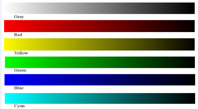
Each of the six dataset files included a random assortment of 30 calcifications obtained from the phantom images and sorted into groups of six according to their diameters. Each dataset file was displayed in one of the six color scales: gray, red, green, yellow, blue, and cyan by using an open-source software (image-J; National Institutes of Health, Bethesda, Md). Each color scale is shown in (Figure 2). The phantom images displayed in each color scale are shown in (Figure 3). The color scale in which each image was presented was random for every observer. The observers were ten radiologists with more than 10 years of experience in interpreting mammograms. None of the observers were color-blind. The radiologists identified the number of differentially sized calcifications, each of which belonged to one of five classes of calcification. The observer was able to coordinate the window width and level at any time. It was assumed that the illuminance of the observation room was constant, and the order in which the colors were assessed was random. The observation of the 30 pieces presented in the six different colors took an average of 70 minutes.
Figure 2: The look-up table of gray, red, yellow, green, blue, and cyan. The look-up table associates saturation with the signal intensity; the highest saturation presented at the left extreme of the scales indicates the highest signal intensity.
Figure 3: The displayed phantom images in each color scale.
III Detection Index Point
Phantom calcifications were generated in five groups of six; the observer evaluation scores were defined by the sum of the products of the identified calcifications in a given group and 0.166. For example, if all six calcifications in all five groups were identifiable, the observers would achieve a score of 5 (6*0.166*5); however, if all six calcifications were identifiable in four of the groups, and only three calcifications were recognizable in the fifth group, the observer would attain a score of 4.5 ([6*0.166*4]+[3*0.166]). Each group of calcifications was observed 10 times by each radiologist; the highest number of calcifications identified in a given trial was retained for the calculation of the detection index.
Figure 4: Box-and-whiskers plots of the detectability achieved by the 10 observers at each color scale. The detection was significantly higher in the yellow scale than the gray (P < 0.05), cyan (P < 0.05), green (P < 0.05), blue (P < 0.01), and red scales (P < 0.01).
Figure 5: Cyan induced less fatigue than yellow (P < 0.05). The fatigue induced by yellow and the gray were not significantly different; both induced significantly less fatigue than green (P < 0.01) red (P < 0.001), and blue (P < 0.001).
IV Fatigue at Observation
After the observation experiment, eyes fatigue was evaluated for the six colors on a six-point scale: a score of "6" indicated little fatigue, while a score of "1" indicated the most. It was assumed that the fatigue corresponded to both visual and psychological fatigue. Each of the ten observers submitted fatigue evaluations.
V Statistical Analysis
The Kruskal–Wallis one-way analysis of variance was performed to determine differences in the detectability of calcifications and the induced eye fatigue among the different color scales. Differences were confirmed with the Mann–Whitney U test. P-values of < 0.05 were considered to indicate statistical significance.
Results
Differences in the detectability of calcifications by the ten observers according to color are shown in (Figure 4). The detectability achieved with yellow was significantly higher than those attained with the other colors: yellow vs. red or blue, P < 0.01; yellow vs. gray, green, or cyan, P < 0.05. Results concerning induced fatigue are shown in (Figure 5). Cyan induced the significantly less fatigue in the 10 observers than did yellow or grey (p < 0.05). No significant difference was observed between the fatigue induced by yellow- and gray-scale displays. Gray was associated with less fatigue than green (p < 0.01), red, and blue (p < 0.001).
Discussion
The visual response of human eyes occurs when photoreceptor cells are excited by light while the responses of neighboring cells are suppressed. This phenomenon is referred to as "contour visual characteristics of the eye," which entails lateral inhibition and the consequent improvement of contrast distinction. The capacity of the human eye to detect objects differs according to the wavelength of light registered – i.e., the color of the perceived object. The colors to which human photopic vision is the most sensitive and, hence, that enable the most accurate visual discrimination are green and yellow in a luminous efficiency function [16-19]. These results agree with the literature [14, 15]. However, we speculated that the characteristics of vision may also differ according to contrast. The present study therefore compared the effect of different color scales on the detectability of microcalcifications at low contrast. We found that visualizing mammograms in yellow scale achieved superior resolution and distinction at low contrast. Taken in the context of previous findings, the detectability of both low-contrast and minute high-contrast lesions may benefit from the use a yellow-scale display rather than gray-scale images, which are currently the most common in digital diagnostic assessments. Clinicians responsible for assessing mammograms are exposed to monitors for extended periods throughout the day; reducing the eye strain consequent of their working conditions is thus of vital consideration to the improvement of the diagnostic assessment of mammograms. Cyan induced the least amount of eye fatigue, while that caused by yellow- and gray-scale displays did not differ significantly. However, this fatigue may change with habituation. Research on the human psychology of color, reports that yellow prompts feelings of strain and excitement [20]. It may be effective for a clinician to cycle through color-scales to prevent fatigue and promote mental stimulation.This study was subject to the important limitation of not having assessed the effect of color-scale on eye fatigue over a long period. Future studies should examine the association between eye fatigue and display color over time.
Conclusion
The present study assessed the effects of different color scales used during digital diagnosis on the detectability of microcalcifications on mammography. Our results indicate that the detectability of the microcalcifications can be improved by using yellow-scale images instead of the more commonly employed grey-scale displays. Concerning eye fatigue, while cyan induced the least amount of fatigue, no difference was found in this respect between yellow- and gray-scale displays.
Conflicts of interest
All authors of this manuscript declare no relationship with any company whose products or services may be related to the subject matter of article.
Funding
All authors state that this work has not received any funding.
Article Info
Article Type
Original ArticlePublication history
Received: Thu 07, Nov 2019Accepted: Tue 26, Nov 2019
Published: Mon 23, Dec 2019
Copyright
© 2023 Akio Ogura. This is an open-access article distributed under the terms of the Creative Commons Attribution License, which permits unrestricted use, distribution, and reproduction in any medium, provided the original author and source are credited. Hosting by Science Repository.DOI: 10.31487/j.RDI.2019.04.04
Author Info
Akio Ogura Fumie Maeda Haruyuki Watanabe Norio Hayashi Toru Negishi
Corresponding Author
Akio OguraGraduate School, Gunma Prefectural College of Health Sciences
Figures & Tables


References
- Fitzmaurice C, Akinyemiju TF, AI Lami FH Alam T, Alizadeh-Navaei R et al. (2018) Global, regional, and national cancer incidence, mortality, years of life lost, year lived with disability, and disability adjusted life-years for 20 cancer groups, 1990-2016: A systematic analysis for the global burden of disease study. JAMA Oncol 4: 1553-1568. [Crossref]
- ACR BI-RADS ATLAS 5th edition 2013, American College of Radiology.
- Scimeca M, Giannini E, Antonacci C, Pistolese CA, Spagnoli LG et al. (2014) Microcalcifications in breast cancer: an active phenomenon mediated by epithelial cells with mesenchymal characteristics. BMC cancer 14: 286. [Crossref]
- Bluekens AM, Holland R, Karssemeijer N, Broeders MJ, den Heeten GJ et al. (2012) Comparison of digital screening mammography and screen-film mammography in the early detection of clinically relevant cancers: a multicenter study. Radiology 265: 707-714. [Crossref]
- Gennaro G, Toledano A, di Maggio C, Baldan E, Bezzon E et al. (2010) Digital breast tomosynthesis versus digital mammography: a clinical performance study. Eur Radiol 20: 1545-1553. [Crossref]
- Spangler ML, Zuley ML, Sumkin JH, Abrams G, Ganott MA et al. (2011) Detection and classification of calcifications on digital breast tomosynthesis and 2D digital mammography: a comparison. AJR Am J Roentgenol 196: 320-324. [Crossref]
- Liu X, Mei M, Liu J (2015) Microcalcification detection in full-field digital mammograms with PFCM clustering and weighted SVM-based method. EURASIP 73: 1-13.
- Wilkinson L, Thomas V, Sharma N (2017) Microcalcification on mammography: approaches to interpretation and biopsy. Br J Radiol 90: 20160594. [Crossref]
- Sickles EA, Brest calcifications: Mammographic evaluation (1986) Radiology 160: 289-293. [Crossref]
- Morgan MP, Cooke MM, McCarthy GM (2005) Microcalcifications associated with breast cancer: an epiphenomenon or biologically significant feature of selected tumors? J Mammary Gland Biol Neoplasia 10: 181-187. [Crossref]
- Weigel S, Decker T, Korsching E, Hungermann D, Böcker W et al (2010) Calcifications in digital mammographic screening: improvement of early detection of invasive breast cancers? Radiology 255: 738-745. [Crossref]
- Murphy WA, DeSchryver-Kecskemeti K (1978) Isolated clustered microcalcifications in the breast: Radiologic-pathologic correlation. Radiology 127: 335-341. [Crossref]
- Spangler ML, Zuley ML, Sumkin JH, Abrams G, Ganott MA et al. (2011) Detection and classifications on digital breast tomosynthesis and 2D digital mammography: a comparison. AJR Am J Roentgenol 196: 320-324. [Crossref]
- Ogura A, Kamakura A, Kaneko Y, Kitaoka T, Hayashi N et al. (2017) Comparison of grayscale and color-scale renderings of digital medical images for diagnostic interpretation. Radiol Phys Technol 10: 359-363. [Crossref]
- Ogura A, Hayakawa K, Tsushima Y, Maeda F, Kamakura A et al. (2018) The yellow scale is superior to the gray scale for detecting acute ischemic stroke on a monitor display in computed tomography. Acad Radiol 25: 1178-1182. [Crossref]
- Wu BW, Fang YC (2015) Human vision model in relation to characteristics of shapes for the Mach band effect. Appl Opt 54: E181-187. [Crossref]
- Schubert EF, Human eye sensitivity and photometric quantities, Light Emitting Diodes.org chapter16.
- Sharpe LT, Stockman A, Jagle W, Jägle H (2005) A luminous efficiency function, V*(λ), for daylight adaption. J Vis 5: 948-968. [Crossref]
- Wright WD (1941) The sensitivity of the eye to small colour differences, Proceedings of the Physical Society 53: 93-112.
- Moutoussis K (2015) The physiology and psychophysics of the color-form relationship: a review. Front Psychol 6: 1407. [Crossref]
